Faculty of Biotechnology October University for Modern Sciences and Arts. Dr. Tarek Yehia Kapiel. B.Sc., M.Sc., Ph.D.
|
|
|
- Shonda Strickland
- 5 years ago
- Views:
Transcription


1
 http://www.msa.")
2 Faculty of Biotechnology October University for Modern Sciences and Arts CELL AND TISSUE CULTURE BT 202 LABORATORY MANUAL Dr. Tarek Yehia Kapiel B.Sc., M.Sc., Ph.D. Faculty of Biotechnology October University for Modern Sciences and Arts (MSA)
3 CELL AND TISSUE CULTURE BT 202 LABORATORY MANUAL CONTENTS CELL AND TISSUE CULTURE TECHNIQUES i PAGE Introduction: 1 What conditions do plant cells need to multiply in vitro? 4 (1). Freedom from competition 4 (2). Nutrients, proper hormones, and removal of waste products 4 (3). A Controlled Environment 5 Why is Tissue Culture Done? 7 BASIC LABORATORY REQUIREMENTS FOR TISSUE CULTURE 9 General Organization 9 Glassware 9 High-purity Water 9 Plant Material 9 Explant 10 PREPARATION OF NUTRIENT MEDIA 11 Nutrient Media for Plant Tissue Cultures 11 I. MINERAL ELEMENTS 12 A. Macroelements consist of N, K, P, Ca, Mg, and S Nitrogen (N) Potassium (K) Phosphorous (P) Magnesium (Mg) Sulfur (S) 14 B. Micronutrients Iron (Fe) Manganese (Mn) Zinc (Zn) 15
4 4. Boron (B) Copper (Cu) Molybdenum (Mo) Cobalt (Co) Iodine (I) Other elements 16 II. ORGANIC COMPOUNDS 16 A. Sugars 16 B. Vitamins 17 C. Myo-inositol 17 D. Complex organics 17 III. ACTIVATED CHARCOAL 18 IV. SOLIDIFYING AGENTS 18 A. Agar 18 B. Agarose 19 C. Gelrite 19 D. Phytagel 20 E. Other supports 20 MEDIA FORMULATIONS 21 PREPARING STOCK SOLUTIONS 23 Macronutrients 24 Micronutrients 24 Vitamins 24 Growth Regulators 25 Storage of Stock Solutions 25 MEDIA PREPARATION 26 Weighing Chemicals 26 Media Preparation from Basal Salt Solutions 27 Media Preparation from Powdered Media 28 Preparation and Use of Vitamin Mixtures 29 VITAMIN SOLUTIONS 30 BASIC LABORATORY PROCEDURES INVOLVED IN MEDIA MAKING: 30 I. Medium Stock Solutions 30 II. Prepared Mixes 31 III. Organic Addenda 31 IV. Making Stock Solutions 32 UNITS USED IN SOLUTIONS 32 ii
5 Volume metrics: 33 Molar concentrations 33 Calculating dilutions of stock solutions: 33 Molecular weights and conversions 34 V. Weighing 36 VI. Measuring Liquids 36 VII. Water 37 VIII. ph 37 IX. Pouring and Storing Media 38 X. Cleaning Glassware 38 STERILE (ASEPTIC) TECHNIQUE 39 I. CONTAMINANTS 40 A. Bacteria, fungi, and insects Bacteria Fungi Yeast Viruses, etc Insects 41 B. Initial Contaminants 41 C. Latent Contamination 42 D. Introduced Contamination 42 E. Detection of Contaminants 42 II. THE TRANSFER HOOD 43 III. STERILIZATION AND USE OF SUPPLIES AND EQUIPMENT: 44 A. Sterilizing tools, media, vessels etc Autoclaving Autoclaving and Fiter-sterilizing Media and Other Liquids Ethylene Oxide Gas UV Radiation Microwave More Comments 46 IV. WORKING IN THE TRANSFER HOOD: 47 V. SURFACE-STERILIZING PLANT MATERIAL Preparation of Stock Plants Ethanol (or Isopropyl Alcohol) Sodium Hypochlorite 52 iii
6 4. Calcium Hypochlorite Mercuric Chloride Hydrogen Peroxide Enhancing Effectiveness of Sterilization Procedure Rinsing Use of Antibiotics and Fungicides in Vitro Plant Preservative Mixture 53 Sterilizing Glassware and Instruments 54 Sterilizing Nutrient Media 55 LAB SAFETY 58 EXPERIMENT 1: Fast tissue culture with rapid-cycling Brassica rapa 60 EXPERIMENT 2: The Effect of Sugar on the Growth of Root Explants 63 EXPERIMENT 3: 63 Production of Plantlets from Floral Organs of Cauliflower 66 EXPERIMENT 4: Callus Formation and Multiplication 75 EXPERIMENT 5: Studies on Carrot Callus Cultures 79 EXPERIMENT 6: Demonstration of "in vitro" Morphogenesis and Totipotency of Seedling Explants 95 EXPERIMENT 7: Effects of Hormone Balance on Explant Growth and Morphogenesis 104 EXPERIMENT 8: Control of organogenesis in cultures of Nicotiana tabacum 114 EXPERIMENT 9: Control of organogenesis in cultures of petals of Saintpaulia ionatha (African violet) 121 EXPERIMENT 10: Establishment of Suspension Cultures 127 EXPERIMENT 11: Anther Culture 129 iv
7 EXPERIMENT 12: Plant Protoplasts Culture 131 EXPERIMENT 13: Growing Agrobacterium cultures 134 v
8 CELL AND TISSUE CULTURE TECHNIQUES INTRODUCTION Cell and tissue culture techniques are essential to many types of academic inquiry, as well as to many applied aspects of biological sciences. In the past, plant tissue culture techniques have been used in academic investigations of totipotency and the roles of hormones in cytodifferentiation and organogenesis. Currently, tissue-cultured plants that have been genetically engineered provide insight into plant molecular biology and gene regulation. Plant tissue culture, the growth of plant cells outside an intact plant, is a technique essential in many areas of the plant sciences. Cultures of individual or groups of plant cells, and whole organs, contribute to understanding both fundamental and applied science. It relies on maintaining plant cells in aseptic conditions on a suitable nutrient medium. The culture can be sustained as a mass of undifferentiated cells for an extended period of time, or regenerated into whole plants. Designing a strategy to culture cells from a plant for the first time can still seem like a matter of trial and error, and luck. However, the commercial production of valuable horticulture crops by micropropagation, which relies on tissue culture, shows that it exists in the routine, as well as experimental, world. Plant tissue culture techniques are also central to innovative areas of applied plant science, including plant biotechnology and agriculture. For example, select plants can be cloned and cultured as suspended cells from which plant products can be CELL AND TISSUE CULTURE TECHNIQUES 1
9 harvested. In addition, the management of genetically engineered cells to form transgenic whole plants requires tissue culture procedures; tissue culture methods are also required in the formation of somatic haploid embryos from which homozygous plants can be generated. Thus, tissue culture techniques have been, and still are, prominent in academic and applied plant science. Plants have been vegetatively propagated for a very long time. Separating rootstocks, grafting, rooting branches and leaves are all ways to vegetatively propagate plants, bypassing the seed stage. Tissue culture is a newer method that enables more control of environmental factors and has provided the evidence that entire, fertile, seed-producing plants can be cloned from single somatic cells. Tissue culture is the method that begins the process for making genetically engineered plants through recombinant DNA technology. However, for decades before recombinant DNA technology started being applied to plants, plant cultures were genetically modified using mutagenic chemicals like colchicine, which often generated larger plants with multiple sets of chromosomes (polyploidy) or by treatment with X-rays to induce mutations via physical breaks in the chromosomes, translocations or changes in the nucleotide sequence (though the mechanisms of mutation at the molecular level were not known at the time these treatments were being used). Plant tissue cultures can be grown in agar medium or liquid medium. On agar, a solid substrate, the plants can more easily develop roots and shoots. In suspension culture, the plant material is generally shaken continuously though gently. Bits and pieces break off starting new clumps. These clumps can be pipetted onto an agar surface or the clumps can be coated with a variety of materials. Seed potatoes are actually clumps of suspensioncells. CELL AND TISSUE CULTURE TECHNIQUES 2
10 Using plant parts (explants) scientists now study the nutritional and regulator requirements for plant development and cell differentiation and for determining how plants respond to their environment at the molecular level, including how plant cells defend themselves from pathogens (fungi, bacteria, viruses). Many common food crops and household plants are vegetatively propagated through grafting or tissue culture. Grapes, seedless fruit and roses are generally grafts. Potatoes, African violets, asparagus fern are routinely propagated from tissue cultures. In fact, 95% of the potatoes we eat are generated from seed embryos of apical meristem suspension cultures. Depending on the plant, tissue cultures might be produced from any of these parts: Apical meristem Flowers Ovary Pollen Stem Leaf Root, and even Seed. Generally, dicots have been more successfully tissue cultured, but recently monocots like rice have been put into culture. Cell and tissue culture is a VERY long-term effort, because it takes weeks for the cultures to grow. However, set-up by an entire class will take only 1 or 2 class sessions, Data collection (observations and measurements) can occur during class breaks. CELL AND TISSUE CULTURE TECHNIQUES 3
11 What conditions do plant cells need to multiply in vitro? Plant cells can be grown in isolation from intact plants in tissue culture systems. The cells have the characteristics of callus cells, rather than other plant cell types. These are the cells that appear on cut surfaces when a plant is wounded and which gradually cover and seal the damaged area. Pieces of plant tissue will slowly divide and grow into a colourless mass of cells if they are kept in special conditions such as: (1). Freedom from competition Many early tissue culture experiments failed, at least in part, because they were not maintained in sterile conditions. Isolated fragments of a plant are extremely disadvantaged in comparison to pathogenic competitors that are complete and unhindered, in reality flourishing, in a culture environment. Bacteria, fungi, and other organisms which can be resisted to some degree by a whole plant can easily outcompete an isolated fragment of tissue from the plant in the relatively nutrient-rich environment of a culture flask. Therefore it is necessary to remove competitor organisms from the culture and isolate it in aseptic conditions. This is usually done by chemical surface sterilization of the explant with an agent such as bleach at a concentration and for a duration that will kill or remove pathogens without injuring the plant cells beyond recovery. The medium and culture flasks used must also be sterile. (2). Nutrients, proper hormones, and removal of waste products When a small portion of a plant is isolated, it is no longer able to receive nutrients or hormones from the plant, and these must be provided to allow growth in vitro. The CELL AND TISSUE CULTURE TECHNIQUES 4
12 composition of the nutrient medium is for the most part similar, although the exact components and quantities will vary for different species and purpose of culture. Types and amounts of hormones vary greatly. In addition, the culture must be provided with the ability to excrete the waste products of cell metabolism. This is accomplished by culturing on or in a defined culture medium, which is periodically replenished. (3). A Controlled Environment Tissue cultures, sustained by the nutritive medium and confined in a protective vessel, require a stable and suitable climate. Thus light and temperature must be more carefully regulated than would be the case for a whole plant. The plant cells can grow on a solid surface as friable, palebrown lumps (called callus), or as individual or small clusters of cells in a liquid medium called a suspension culture. These cells can be maintained indefinitely provided they are sub-cultured regularly into fresh growth medium. Tissue culture cells generally lack the distinctive features of most plant cells. They have a small vacuole, lack chloroplasts and photosynthetic pathways and the structural or chemical features that distinguish so many cell types within the intact plant are absent. They are most similar to the undifferentiated cells found in meristematic regions which become fated to develop into each cell type as the plant grows. Tissue cultured cells can also be induced to redifferentiate into whole plants by alterations to the growth media. Plant tissue cultures can be initiated from almost any part of a plant. The physiological state of the plant does have an influence on its response to attempts to initiate tissue culture. The parent plant must be healthy and free from obvious signs of disease or decay. The source, termed CELL AND TISSUE CULTURE TECHNIQUES 5
13 explant, may be dictated by the reason for carrying out the tissue culture. Younger tissue contains a higher proportion of actively dividing cells and is more responsive to a callus initiation programme. The plants themselves must be actively growing, and not about to enter a period of dormancy. The exact conditions required to initiate and sustain plant cells in culture, or to regenerate intact plants from cultured cells, are different for each plant species. Each variety of a species will often have a particular set of cultural requirements. Despite all the knowledge that has been obtained about plant tissue culture during the twentieth century, these conditions have to be identified for each variety through experimentation. Plant tissue culture now has direct commercial applications as well as value in basic research into cell biology, genetics and biochemistry. The techniques include culture of cells, anthers, ovules and embryos on experimental to industrial scales, protoplast isolation and fusion, cell selection and meristem and bud culture. CELL AND TISSUE CULTURE TECHNIQUES 6
14 Why is Tissue Culture Done? Tissue culture offers numerous significant benefits over traditional propagation methods: Propagation can be much more rapid than by traditional means. Large numbers of genetically identical clones may be produced. Micropropagation using meristem and shoot culture to produce large numbers of identical individuals. It may be possible in vitro to multiply plants that are very difficult to propagate by cuttings or other traditional methods Seeds can be germinated with no risk of damping off/predation. Under certain conditions, plant material can be stored in vitro for considerable periods of time with little or no maintenance. Tissue culture is an essential part of many genetic transformation protocols. Tissue culture techniques are used for screening programmes of cells, rather than plants for advantageous characters. Tissue culture techniques are used for large-scale growth of plant cells in liquid culture as a source of secondary products Tissue culture techniques are used for crossing distantly related species by protoplast fusion and regeneration of the novel hybrid Tissue culture techniques are used for production of dihaploid plants from haploid cultures to achieve homozygous lines more rapidly in breeding programmes. Tissue culture techniques are used for virus eradication, genetic manipulation, somatic hybridization and other procedures that benefit propagation, plant improvement, and basic research. The processes studied in the experiments which follow have been widely used industrially. They are summarized in the flow diagram (Figure 1). CELL AND TISSUE CULTURE TECHNIQUES 7
15 Figure 1. Flow chart summarizing Plant cell and tissue culture experiments. CELL AND TISSUE CULTURE TECHNIQUES 8
16 BASIC LABORATORY REQUIREMENTS FOR TISSUE CULTURE General Organization Localize each portion of the tissue culture procedure in a specified place in the laboratory. An assembly-line arrangement of work areas (such as, media preparation, glassware washing, sterilization, microscopy, and aseptic transfers) facilitates all operations and enhances cleanliness. Glassware Use glassware that has only been used for tissue culture and not other experiments. Toxic metal ions absorbed on glassware can be especially troublesome. Wash glassware with laboratory detergent, then rinse several times with tap water and, finally, rinse with purified water. High-purity Water Use only high-purity water in tissue culture procedures. Double glass distilled water or deionized water from an ion-exchanger are acceptable. Water should not be stored, but used immediately. Regular maintenance and monitoring of water purification equipment are necessary. Purified water for tissue culture can also be purchased. Plant Material Plants used in tissue culture need to be healthy and actively growing. Stressed plants, particularly water-stressed plants, usually do not grow as tissue cultures. Insect and disease-free greenhouse plants are rendered aseptic more readily, so contamination rate is lower when these plants are used in tissue culture procedures. Seeds that can be easily surface CELL AND TISSUE CULTURE TECHNIQUES 9
17 Explant sterilized usually produce contamination-free plants that can be grown under clean greenhouse conditions for later experimental use. The starting point for all tissue cultures is plant tissue, called an explant. It can be initiated from any part of a plant - root, stem, petiole, leaf or flower - although the success of any one of these varies between species. It is essential that the surface of the explant is sterilized to remove all microbial contamination. Plant cell division is slow compared to the growth of bacteria and fungi, and even minor contaminants will easily over-grow the plant tissue culture. The explant is then incubated on a sterile nutrient medium to initiate the tissue culture. CELL AND TISSUE CULTURE TECHNIQUES 10
18 PREPARATION OF NUTRIENT MEDIA The composition of the growth medium is designed to sustain the plant cells, encourage cell division, and control development of either an undifferentiated cell mass, or particular plant organs. The concentration of the growth regulators in the medium, namely auxin and cytokinin, seems to be the critical factor for determining whether a tissue culture is initiated, and how it subsequently develops. The explant should initially form a callus, from which it is possible to generate multiple embryos and then shoots, forming the basis for plant regeneration and thus the technology of micropropagation. The first stage of tissue culture initiation is vital for information on what combination of media components will give a friable, fastgrowing callus, or a green chlorophyllous callus, or embryo, root or shoot formation. There is at present no way to predict the exact growth medium, and growth protocol, to generate a particular type of callus. These characteristics have to be determined through a carefully designed and observed experiment for each new plant species, and frequently also for each new variety of the species which is taken into tissue culture. The basis of the experiment will be media and protocols that give the desired effect in other plant species, and experience. Nutrient Media for Plant Tissue Cultures One of the first decisions that must be made when developing a tissue culture system is what medium to use. Nutrient media for plant tissue culture are designed to allow plant tissues to be maintained in a totally artificial environment. Many different tissue culture media have been developed, but only a few have found wide-spread use, e.g. MS (Murashige and Skoog, 1962). SH (Shenck and CELL AND TISSUE CULTURE TECHNIQUES 11
19 Hildebrandt), and Gamborg's B5. One of the most successful media, devised by Murashige and Skoog (Murashige and Skoog, 1962) was formulated by analyzing the inorganic components in tobacco plants and then adding them to medium in amounts similar to those found in the plants. Not only did they find that the ions themselves were important, but the forms in which the ions were supplied were critical as well. In addition to mineral elements, the macro- and micronutrients that are similar to what is found in fertilizers, nutrient media also contain organic compounds such as vitamins, plant growth regulators, and a carbon source. I. MINERAL ELEMENTS A. Macroelements consist of N, K, P, Ca, Mg, and S. 1. Nitrogen (N) Nitrogen is essential for plant growth. Most inorganic nitrogen is converted to amino acids and then to proteins. Nitrogen is typically added to plant nutrient media as the nitrate ion (NO 3-, oxidized) and/or the ammonium ion (NH 4+, reduced), which are added as inorganic salts. Inorganic nitrogen generally ranges from mm in nutrient media. In devising media, both the total amount of nitrogen as well as the relative amounts of NO 3- and NH 4+ are important. There are usually lower levels of NH 4+ than NO 3- in medium; nitrate is usually added at concentrations between 25 and 20 mm and ammonium at concentrations between 2 and 20 mm. For example, the amount of NH 4+ in MS medium is less than half that of NO 3- and in other media the NH 4+ concentration is lower still. Cultures of some species can proliferate on medium containing nitrates alone, CELL AND TISSUE CULTURE TECHNIQUES 12
20 and some can grow on a medium with ammonium as the sole inorganic nitrogen source if one or more of the TCA cycle acids (citrate, succinate, malate) are included in the medium at concentrations of about 10 mm. In poorly buffered media, the use of both nitrogen forms helps maintain ph. Also, many plant species appear to respond best if they are given both forms, although the reason for this is not known. Nitrogen may also be added to medium in an organic form, as amino acids such as proline or glutamine, hydrolysates (such as casein hydrolysate), or, as above, as organic acids. Organic nitrogen is already reduced, i.e. in the form in which most nitrogen exists in the plant, and so may be taken up more readily than inorganic nitrogen. The organic forms are often added to media that do not contain ammonium. However, almost always, some inorganic nitrogen is present. 2. Potassium (K) Potassium is the major ion in plants with a positive charge, balancing negative ions. Although the amount of potassium required varies widely among different species, in media potassium concentration is generally correlated with that of nitrate and ranges between mm. 3. Phosphorous (P) Phosphorus is an integral part of nucleic acids and other structural compounds. It is added to culture medium as phosphate (PO 4- ) in sodium or potassium hydrogen phosphates in concentrations ranging from 1-3 mm. 4. Calcium (Ca) Calcium is a co-factor of many enzymes and is particularly important in cell wall synthesis. It is supplied mostly as calcium chloride or calcium nitrate, concentrations ranging CELL AND TISSUE CULTURE TECHNIQUES 13
21 between 1 and 3 mm. In plant cultures, calcium deficiency may result in shoot tip necrosis. 5. Magnesium (Mg) Magnesium is critical for the functioning of enzymes, is an integral component of the chlorophyll molecule, and is a cation that balances negative ions. It is usually added as magnesium sulfate in concentrations similar to that of calcium. 6. Sulfur (S) Sulfur is a part of several amino acids and has an important function in protein structure. It is supplied as the SO 4- ion, generally with magnesium as the cation, in concentrations ranging from 1-3 mm. B. Micronutrients Micronutrients used in plant tissue culture are Fe, Mn, Zn, B, Cu and Mo, Co, and I. 1. Iron (Fe) Iron is necessary for chlorophyll synthesis and functions in many oxidation/reduction reactions. It is generally present in media at approximately 1 M. The major problem in supplying iron in vitro is that it forms insoluble compounds in alkaline ph, a problem that is particularly evident in liquid culture, where it may be seen as a precipitate. The use of chelating agents, which bind metal ions, makes iron more stable and available to plant tissues over wider ph ranges. Although there are several of these, the sodium or potassium form of ethylenediaminetetraacetic acid (EDTA) is most often used because it is not as toxic as other chelating agents and it enables iron to be available to cultures over a wider ph CELL AND TISSUE CULTURE TECHNIQUES 14
22 range than other agents. Fe-EDTA may be purchased as a salt or prepared from ferric sulfate and EDTA. 2. Manganese (Mn) Manganese is required for enzyme reactions, particularly in respiratory and photosynthetic processes and is usually added as manganese sulfate in concentrations of 5-30 M. 3. Zinc (Zn) Zinc is also required in many enzyme activities and is added to medium in concentrations similar to that of manganese. The most common form in which zinc is added is as the sulfate salt. 4. Boron (B) Boron is an essential element involved in lignin biosynthesis and metabolism of phenolic acids and is supplied as boric acid in culture medium ( M). Boron deficiency results in the death of shoot tip meristems. 5. Copper (Cu) Copper is critical in many enzyme reactions, including the cytochrome oxidase system. It is added to culture medium (as cupric sulfate) in very low concentrations (0.1 M), because high amounts can be toxic. 6. Molybdenum (Mo) Molybdenum functions in the transformation of nitrate to ammonium. It is added as sodium molybdate in low concentrations (1 M) in culture medium. CELL AND TISSUE CULTURE TECHNIQUES 15
23 7. Cobalt (Co) Cobalt is not considered to be an essential mineral by plant physiologists, but is included in many of the most widely used media formulations. Cobalt is supplied in concentrations similar to that of copper, again because it may be toxic at higher concentrations. 8. Iodine (I) Iodine is not considered to be an essential element, but it is often added to plant culture media (5 M) because it has been found to improve growth of roots and callus in vitro. 9. Other elements Nickel (Ni), aluminum (Al), and silicon (Si), are added to a few media formulations. These elements have not been found to be necessary for most plant species in vitro. II. ORGANIC COMPOUNDS Organic compounds are also added to plant culture medium. Some of these compounds, such as sugars, are absolutely needed for growth, while others, such as vitamins, undefined compounds, and organic acids, may not be essential but may enhance growth. A. Sugars Most plant tissue cultures are not highly autotrophic, that is, capable of fixing carbon through photosynthesis, due to limitations in culture of CO 2 availability, among other factors. Therefore, sugar is added to the medium as an energy source. Sucrose is the most common sugar added, although glucose, fructose, and sorbitol are also used in certain instances. Sucrose is the sugar form most commonly transported in plants; it is broken down into glucose and CELL AND TISSUE CULTURE TECHNIQUES 16
24 fructose during metabolism. It is also partially hydrolyzed into glucose and fructose during autoclaving. The concentration of sugars in nutrient media generally ranges from 20 to 40 g/l. Sugars also act as an osmoticum in the medium. Osmotic potential can have an important effect on in vitro response. Nutrient salts contribute from 20% to 50% of the osmotic potential of media, with sucrose making up the rest. When sucrose is hydrolyzed, as during autoclaving, its contribution to the osmotic potential is further increased. B. Vitamins Only thiamine (vitamin B1), which is required for carbohydrate metabolism and the biosynthesis of some amino acids, has been shown to be essential for most plant cultures. Nicotinic acid (niacin) and pyridoxine (B6) are also commonly added to Murashige and Skoog medium and some other media. Other vitamins such as biotin, folic acid, ascorbic acid (vitamin C), and vitamin E (tocopherol) are sometimes added to media formulations. Vitamin concentrations are generally very low. C. Myo-inositol Myo-inositol, a sugar alcohol, is added to most plant culture media. Although not essential for culture viability, it can significantly improve in vitro response, especially in monocots. Although myo-inositol is not essential for growth of many plant species, its effect on growth is significant. D. Complex organics These are undefined supplements such as coconut milk, coconut water, yeast extract, fruit juices and fruit pulps. They may supply amino acids, vitamins, plant growth regulators, and/or secondary plant metabolites. Complex organics were CELL AND TISSUE CULTURE TECHNIQUES 17
25 frequently used early in the history of plant tissue culture, when growth requirements were less defined. Now they are used only when no combination of defined components supports growth. Their disadvantages are that the important compounds in them are not known, and may vary greatly from batch to batch. Only protein hydrolysates and coconut milk (at 5-20% v/v) are used much today. III. ACTIVATED CHARCOAL Activated charcoal is sometimes added to media in order to adsorb toxic compounds released by plant tissues, particularly oxidized phenolics. It may be especially useful in rooting medium. However, activated charcoal adsorbs not only toxic compounds, but also growth regulators and other compounds that are added to the medium. Activated charcoal is usually acid-washed prior to addition to the culture medium at a concentration of %. IV. SOLIDIFYING AGENTS A. Agar Solidifying agents are used to create semi-solid or solid media wherein plant cultures are not submerged in the medium. Liquid medium can be used for many plants but it must usually be agitated to provide sufficient oxygen to the tissue. Agar is the most commonly used gelling agent. Marine red algae contain the structural polysaccharide agar, which consists of 2 components, agarose and agaropectin. Agarose is composed of alternating D-galactose and 3,6- anhydro-l-galactose with side chains of 6-methyl-Dgalactose residues. Agaropectin is like agarose but additionally contains sulfate ester side chains and D- glucuronic acid. The tertiary structure of agarose is a double helix with a so-called threefold screw axis. The central cavity CELL AND TISSUE CULTURE TECHNIQUES 18
26 of this double helix can accommodate H2O molecules. Agarose and agaropectin readily form gels that contain high amounts of H2O (up to 99.5%). When agar is mixed with liquid, it forms a gel that melts at about 100 C and solidifies at about 45 C. Other benefits are that agar does not react with any components of the medium and it is not digested by enzymes from the plant tissue. All agar contains impurities, such as inorganic salts, organic compounds, phenolics, and long chain fatty acids; amounts and types vary depending on the manufacturer. These compounds usually do not interfere with culture response. If necessary, agar can be washed to remove inhibitory impurities. Agar does not gel well under acidic conditions (ph <4.5). The inclusion of activated charcoal in media may also inhibit gelling of agar. The agar concentrations commonly used in plant culture media range between 0.5% and 1%; these concentrations yield a firm gel at typical media phs. The concentration of agar may be critical to plant response in culture. Medium that is too soft may produce hyperhydric (abnormal, glassy-looking) tissue while medium that is too hard may cause reduced growth. B. Agarose When greater purity is needed, agarose may be used. Agarose is extracted from agar leaving behind agaropectin and its sulfate groups. Because of the additional purification, agarose is considerably more expensive than agar. Agarose also has higher gel strength than agar and thus less is required for solidification of media. Agarose is used in situations where the impurities of agar are a major Disadvantage, such as in protoplast culture. C. Gelrite Gelrite consists of a polysaccharide produced by the bacterium Pseudomonas elodea. Medium solidified with Gelrite has the advantage of being clear, which agar- CELL AND TISSUE CULTURE TECHNIQUES 19
27 solidified medium is not. Consequently contamination is more easily detected at an early stage. Impurities in Gelrite contain inorganic ions, but no organic compounds. Gelrite requires more stirring than agar when being added to media. Unlike agar, Gelrite cannot be reheated and gelled successfully. One limitation of Gelrite is that the concentration of divalent cations such as calcium and magnesium ions must be within the range of 4-8 mm/liter. Concentrations of these two ions either less than or greater than this range result in the media not gelling. Gelrite may also produce hyperhydric plants when used at low concentrations. D. Phytagel Phytagel is an agar substitute produced from a bacterial substrate composed of glucuronic acid, rhamnose and glucose. It produces a clear, colorless, high-strength gel, which aids in detection of microbial contamination. Phytagel provides an economical alternative to agar as a gelling agent. It is used at a concentration of g/l. To prevent clumping, Phytagel should be added to rapidly stirring culture medium which is at room temperature. Hyperhydricity may also be a problem with this gelling agent. The selection of a gelling agent for specific plants is generally empirical. For unknown reasons, tissues of some species grow more vigorously on one gelling agent than on another. Another major consideration is the degree of hyperhydricity induced in a species by the different gelling agents. One potential way to overcome this is to combine agar and either Gelrite or Phytagel in the medium. E. Other supports Mechanical supports such as filter paper folded into wicks and polyethylene rafts can be used with liquid medium to CELL AND TISSUE CULTURE TECHNIQUES 20
28 ensure an adequate supply of oxygen. Many other materials have been used as well including rock wool, cheesecloth, sand, and pieces of foam. Explants can be grown in rotating cultures where the tissue is alternately bathed in liquid and exposed to air. MEDIA FORMULATIONS Although the most used medium formulation is that of Murashige and Skoog (1962) (references are in the Gamborg paper), many others have been developed. Murashige and Skoog (MS) medium was developed for culture of tobacco and was formulated based on an analysis of the mineral compounds present in the tobacco tissue itself. It has comparatively high salt levels, particularly of K and N. Linsmaier-Skoog medium (Linsmaier and Skoog, 1965) is a version of MS medium with modified organic constituents. White's medium (White, 1963), a low salt formulation, was developed originally for the culture of tomato roots. Gamborg's B5 medium (Gamborg et al., 1968) was developed for soybean callus culture and contains a much greater proportion of nitrate compared to ammonium ions. The vitamins in this medium formulation are also often added to MS salts. Schenk and Hildebrandt (1972) developed their medium for the culture of callus of both monocots and dicots. Nitsch and Nitsch (1969) medium was developed for anther culture and contains lower salt concentrations than MS medium, but not as low as in White's medium. Lloyd and McCown's Woody Plant Medium (WPM) (Lloyd and McCown, 1981) has been used successfully for a great many tree species. Knudson's medium has been used in orchard culture (Knudson 1946). CELL AND TISSUE CULTURE TECHNIQUES 21
29 There are several important observations to make about these media: All of them are fully defined (no complex organics). A chelated iron source is preferred (Fe-EDTA). MS and SH are high salt media. MS and SH use both ammonium and nitrate ions as nitrogen sources. SH contains a very high level of myo-inositol. Sucrose is the carbon source. Table 1. Formulations of Common Plant Culture Media * Concentration in culture medium (mg/liter) Constituent MS SH B5 KNO NH4NO NH4H2PO4 300 (NH4)2SO4 134 MgSO4 7H2O CaCl2 2H2O KH2PO4 170 NaH2PO4 H2O 150 MnSO4 H2O MnSO4 4H2O 22.3 KI H3BO ZnSO4 7H2O CuSO4 5H2O Na2MoO4 2H2O CoCl2 6H2O FeSO4 7H2O Na2EDTA Nicotinic acid Pyridoxine-HCl Thiamine-HCl myo-inositol Glycine 2.0 Sucrose CELL AND TISSUE CULTURE TECHNIQUES 22
30 In complex media such as these, consisting of many components, there are a huge number of permutations of substances and concentrations to test to compose an ideal medium for a particular plant species and genotype. When working with a species new to you, the first place to start is in the literature, finding out what other people have used for that species or one closely related. However, there may be considerable differences in requirements for different cultivars. In practice, researchers may test several basal media, but only try to optimize a few components, in particular, plant growth regulators (PGRs). PREPARING STOCK SOLUTIONS The use of stock solutions reduces the number of repetitive operations involved in media preparation and, hence, the chance of human or experimental error. Moreover direct weighing of media components (e.g., micronutrients and hormones) that are required only in milligram or microgram quantities in the final formulation cannot be performed with sufficient accuracy for tissue culture work. For these components, preparation of concentrated stock solutions and subsequent dilution into the final media is standard procedure. In addition, concentrated solutions of some materials are more stable and can be stored for longer periods than more dilute solutions. To prepare a stock solution, weigh out the required amount of the compound and place it in a clean flask. It is common practice to make a stock solution 10x or 100x, depending upon the solubility of the compound. Once the chemical is in the flask, dissolved it in a small amount of water, ethyl alcohol, 1 N NaOH, or 1 N HCL. Next, slowly add doubledistilled water to the flask, while agitating. Continue this until the proper volume is reached. Label the flask with the name of the solution, preparation and expiration dates, and the name of the person who prepared the solution. Certain CELL AND TISSUE CULTURE TECHNIQUES 23
31 items, e.g., IAA, must be prepared and stored in amber bottles to prevent photodecomposition. The volumes of stock solutions prepared at various concentrations that must be used to achieve various final concentrations are presented in tabular form in the Plant Growth Regulator Section. Macronutrients Stock solutions of macronutrients can be prepared at 10 times the concentration of the final medium. A separate stock solution for calcium salts may be required to prevent precipitation. Stock solution of macronutrients can be stored safely for several weeks in a refrigerator at 2-4 C. Micronutrients Micronutrient stock solutions are generally made up at 100 times their final strength. It is recommended that micronutrient stocks be stored in either a refrigerator or freezer until needed. Micronutrient stock solutions could be stored in a refrigerator for up to 1 year without appreciable deterioration. Iron stock solutions should be prepared and stored separately from other micronutrients in an amber storage bottle. Formulations for preparing stock solutions of iron are presented later. Vitamins Vitamins are prepared as 100X or 1000X stock solutions and stored in a freezer (-20 C) until used. Vitamin stock solutions should be made up each time media is prepared if a refrigerator or freezer is not available. Vitamin stock solutions can be stored safely in a refrigerator for 2-3 months but should be discarded after that time. CELL AND TISSUE CULTURE TECHNIQUES 24
32 Growth Regulators The auxins NAA and 2,4-D are considered to be stable and can be stored at 4 C for several months; IAA should be stored at -20 C. Auxin stock solutions are generally prepared at times the final desired concentrations. Solution of NAA and 2,4-D can be stored for several months in a refrigerator or indefinitely at -20 C. Generally IAA and 2,4-D are dissolved in a small volume of 95% ethyl alcohol or KOH and then brought to volume with double-distilled water; NAA can be dissolved in a small amount of 1 N NaOH or KOH, which also can be used to dissolve 2,4-D and IAA. The cytokinins are considered to be stable and can be stored at -20 C. Cytokinin stock solutions are generally prepared at 100X to 1000X concentrations. Many of the cytokinins are difficult to dissolve, and a few drops of either 1 N HCL, 1 N NaOH, in KOH or DMSO, are required to bring them into solution. Storage of Stock Solutions Storage conditions for most stock solutions have already been pointed out; however, some additional points can be made. For convenience, many labs prepare stock solutions and then divide them into aliquots sufficient to prepare from 1 to 10 liter of medium; these aliquots are stored in small vials or plastic bags in a freezer. This procedure removes the inconvenience of having to un-thaw a large volume of frozen stock each time medium is prepared. Some have found that heating in a microwave oven is a satisfactory and quick method of thawing concentrated medium. CELL AND TISSUE CULTURE TECHNIQUES 25
33 MEDIA PREPARATION Weighing Chemicals The preparation of media requires careful weighing of all components. Even if a commercially prepared medium is used, care must be taken in preparing it and any stock solutions that are required. Because of the diversity of laboratory balances in use, it is impossible to review the details of their operation. The manufacturer s instructions should be consulted before using any balance. The types of balances most often encountered in the laboratory include top-loading single-pan balance, triplebeam balance, double-pan torsion balance, analytical singlepan balance, and top-loading electronic balance. The last type has become quite popular in recent years due to its accuracy, ease of use, and durability. With certain models of top-loading electronic balances, milligram accuracy is possible. Such accuracy previously required the use of analytical balances. Several common precautions must be observed to obtain accurate weights. First, the balance should be located on a hard, stable, level surface, which is free of vibrations and excessive air drafts. The balance or weigh area should always be kept clean. Most importantly, the balance should never be overloaded (see manufacturer s specification). It is advisable to use a lightweight weighing container or paper rather than placing the material to be weighted directly on the pan surface. Media Preparation from Basal Salt Solutions Liquid 10X solutions are offered for your convenience. To avoid precipitation over long-term storage, solutions mixed at the proper dilution to make a solution with the CELL AND TISSUE CULTURE TECHNIQUES 26
34 appropriate salt concentration. The basic steps for preparing 1 liter of culture medium are listed below. 1. Measure out approximately 700 ml of tissue culture grade water. 2. While stirring the water, add 100 ml of Macronutrient Solution 3. Continue stirring the mixture while adding 100 ml of Micronutrient Solution 4. Add desired heat stable supplements (e.g. sucrose, gelling agent, vitamins, auxins, cytokinins, etc.) 5. Add additional tissue culture grade water to bring the medium to the final volume. 6. While stirring, adjust medium to desired ph using NaOH, HCl or KOH. 7. If gelling agent is used, heat until the solution is clear. 8. Dispense the medium into the culture vessels before (or after) autoclaving according to your application. Add heat labile constituents after autoclaving. 9. Sterilize the medium in a validated autoclave at 1 kg/cm 2 (15 psi), 121 C, for the time period described under Sterilization of Media. 10. Allow medium to cool prior to use. CELL AND TISSUE CULTURE TECHNIQUES 27
35 Media Preparation from Powdered Media Powdered media are extremely hygroscopic and must be protected from atmospheric moisture. If possible the entire contents of each package should be used immediately after opening. Preparing the medium in a concentrated form is not recommended as some salt added to the medium may affect shelf life and storage conditions. The basic steps for preparing the culture medium are listed below: 1. Measure out approximately 90% of the final required volume of tissue culture grade water, e.g. 900 ml for a final volume of 1000 ml. Select a container twice the size of the final volume. 2. While stirring the water add the powdered medium and stir until completely dissolved. 3. Rinse the original container with a small volume of tissue culture grade water to remove traces of the powder. Add to the solution in Step Add desired heat stable supplements (e.g. sucrose, gelling agent, vitamins, auxins, cytokinins, etc.) 5. Add additional tissue culture grade water to bring the medium to the final volume. 6. While stirring, adjust medium to desired ph using NaOH, HCl, or KOH. 7. If a gelling agent is used, heat until the solution is clear. 8. Dispense the medium into the culture vessels before (or after) autoclaving according to your application. Add heat labile constituents after autoclaving. CELL AND TISSUE CULTURE TECHNIQUES 28
36 9. Sterilize the medium in a validated autoclave at 1 kg/cm2 (15 psi), 121 C, for the time period described under Sterilization of Media. 10. Allow medium to cool prior to use. * Heating may be required to bring powders into solution. Preparation and Use of Vitamin Mixtures Powdered vitamin mixtures are hygroscopic and must be protected from atmospheric moisture. The entire contents of each package should be used immediately after opening. The basic steps for preparing 1000X concentrated solutions with vitamin mixtures are listed below: 1. Measure out 70% of the final required volume of deionized-distilled water (e.g. 70 ml for a final volume of 100 ml) 2. While stirring the water add the powdered vitamin mixture. Stir until completely dissolved. Increasing the ph and/or warming solution (35-37 C) may be required. 3. Rinse the original container with a small volume of water to remove traces of the powder. Add to the solution in Step No Add additional water to bring the medium to the final volume. 5. The resulting 1000X concentrated solution should be used at a concentration of 1 ml/l of medium. 6. Follow the same steps to prepare a 100X concentrated solution and use at 10 ml/l of medium. CELL AND TISSUE CULTURE TECHNIQUES 29
37 VITAMIN SOLUTIONS 1. The vitamin solutions are sterile filtered through a double 0.2 μm filtration unit and are ready for use. 2. Vitamin solutions should be added at a concentration of 1 ml/l of medium to prepare the final recommended concentration of vitamins in the medium. BASIC LABORATORY PROCEDURES INVOLVED IN MEDIA MAKING: The majority of laboratory operations utilized in the in vitro propagation of plants can be easily learned. One needs to concentrate mainly on accuracy, cleanliness, and strict adherence to details when performing in vitro techniques. I. Medium Stock Solutions In the old days, mineral salt mixtures were prepared as stock solutions ranging from 10 to 100 times the final concentrations used in the medium. Stock solutions can be prepared as two solutions, one containing all of the macronutrients and one containing all of the micronutrients. These solutions must be kept fairly dilute (10-20X) in order to avoid precipitation of calcium and magnesium phosphates and sulfates. A more common method is to arrange mineral salt stock solutions according to the ions they contain. A series of solutions containing the inorganic components of the medium is prepared; precise combinations may vary from lab to lab. Using this method, salt stocks can be prepared in 100X concentrations. Iron-EDTA chelate is prepared from CELL AND TISSUE CULTURE TECHNIQUES 30
38 iron sulfate and Na-EDTA by mixing the proper amounts of the two compounds with water and then autoclaving. This stock must then be stored in a dark container. The prepared stock solutions are usually stored in the refrigerator. Although the initial cost of chemicals may be substantial, overall the ongoing cost of using stock solutions is probably less than that of using prepared mixes (unless you factor in labor). However, the stocks must be prepared over and over when they are used up, and each time this is done potential error is introduced. It is best if one person consistently prepares the stocks. Most people no longer make up stock solutions of the inorganic medium components. Stocks solutions may be useful if many different media are made in the lab or frequent changes in concentrations are made in individual components. II. Prepared Mixes Several companies sell prepared salt and vitamin mixtures as powders. These are easily handled by adding the proper amount of powder to water. The mixtures can be purchased as complete media or as salts alone. The packs often contain the necessary ingredients for one liter of medium. The contents are extremely hyroscopic once a pack is opened, so it is best to use the contents all at once. A small amount of precipitate may be seen in media prepared using either of these methods. This result from iron being displaced from the chelate over time as media is stored. It doesn t appear to be deleterious. III. Organic Addenda Most organic addenda are added in relatively low concentrations, too low to be weighed out accurately. Therefore, stock solutions ranging from 100 to 1000X final concentrations are prepared. Vitamins may be prepared as CELL AND TISSUE CULTURE TECHNIQUES 31
39 stocks or purchased premade. Vitamin solutions can be divided into aliquots and stored in the freezer at -20 C. Most growth regulators are stable for up to a month in stock solutions. The solvents used for dissolving growth regulators vary depending on the compound under consideration. IV. Making Stock Solutions There are three ways to describe amounts of chemicals in a stock: parts per million (rarely used), as grams per liter, or as molar units (journals usually require concentrations to be expressed this way). UNITS USED IN SOLUTIONS 1. mg/l = milligrams of substance in one liter (1000 ml) of solution. 2. ppm = part per million 1 unit of substance in one million units of solution 1 mg/l = 1 ppm 3. mole = gram molecular weight The formula weight of the substance in grams. FW (formula weight = sum of weights of atoms in the formula) e.g. H2O 1 x = 18 1 mole H2O = 18 grams e.g. sucrose C12H22O11 FW = mole = grams For preparing molar solutions, it is necessary to know molecular weight. To calculate how much of a chemical to weigh out to make a stock solution: (FW/desired molar concentration) x the number of liters desired = amount to be dissolved CELL AND TISSUE CULTURE TECHNIQUES 32
40 Volume metrics: Liter ml 10-3 L l 10-6 L Pl 10-9 L Molar concentrations mm 10-3 L M 10-6 L nm 10-9 L When working in molar units it is often convenient to prepare a stock as, for example, 10 ml of a 10 µm solution. This is easily remembered as 0.1 X the formula (molecular) weight (in mg) per 10 ml solvent. So, for example, to prepare 10 ml of a 10µM solution of IAA, you would weigh 17.5 mg of IAA and add it to 10 ml of solvent. Calculating dilutions of stock solutions: M1= Molarity of stock solution M2 = Molarity of final solution desired V1 = volume of stock solution needed V2 = volume of final solution desired Usually we know all but V1 Use the simple formula: M1*V1=M2*V2 Can be rearranged to be V1=(M2*V2)/M1 CELL AND TISSUE CULTURE TECHNIQUES 33
41 Example: You have a 1M stock of NaCl, and want to make a 10mM solution, in other words a 0.01 M solution, and you need 100 mls of it. So using the formula: V1=(0.01 M*100mL)/1M = 1 ml, i.e. you need 1 ml of the stock, mixed with 99 ml H20 to have 100 mls of solution. When working in mg/l a convenient stock concentration is 1 mg/ml. The stock concentrations can be prepared so that one only needs to add small volumes to media. Therefore, something like alcohol can be used as a solvent in a stock the stock is added in such a low concentration that the solvent is not toxic. To prepare a stock solution, weigh out the required amount of the compound and place it in a clean flask. Once the chemical is in the flask, dissolve it in a small amount of the proper solvent, usually water, ethyl alcohol, 1 N NaOH, or 1 N HCL. Next, slowly add double-distilled water to the flask, while agitating. Continue this until the proper volume is reached. Label the flask with the name of the solution, preparation and expiration dates, and the name of the person who prepared the solution. Molecular weights and conversions Often literature will address concentration as a molar solution (M). This can be confusing for some, because we use mg/l in this class. The conversion between moles and mg/l is not difficult. First I'll go over how to make a molar solution. A 1 molar (1M) solution is simply the molecular weight in grams in 1 liter of solution (usually water). CELL AND TISSUE CULTURE TECHNIQUES 34
42 Table 2. List of some common chemicals and their molecular weights: CHEMICAL MOLECULAR MOLECULAR CHEMICAL WEIGHT WEIGHT Thiamine HCl Biotin BAP iP Inositol Pyridoxine HCl NAA Glycine ,4 - D Zeatin Nicotinic Acid Glucose Kinetin Sucrose Folic Acid Mannitol IAA Sorbitol If one wishes to make a 1 M solution of thiamine, weight out grams and place this in 1 liter of water. Again, it is simply the molecular weight in grams in 1 liter. A 1 millimolar solution (mm) is 1/1000 of 1M. In the case of thiamine, a 1mM would be made by adding grams in 1 liter. To convert Moles to mg/l, do the following: molecular weight X concentration (moles) 1000 = mg/l Example: A 0.5 M solution of BAP is made by adding 112.6g of BAP per liter. Notice we are talking about a half mole, therefore use one half as much in grams - the volume stays the same. In case you need this info: The dilution from one molarity to another follows the relationship: M 1 X V 1 = M 2 X V 2 where M = molarity, V = volume, 1 is the stock solution and 2 is the new solution. If you have a 12.5 M solution and want 100 ml of a 1 M solution, 12.5 X xml = 1M X 100ml CELL AND TISSUE CULTURE TECHNIQUES 35
43 solving for xml = 8ml. So to make the desired solution add 8 ml to 92 ml of water. V. Weighing The preparation of media usually requires careful weighing of at least some components. Even if a commercially prepared medium is used, care must be taken in preparing it and any stock solutions that are required. Sucrose, myoinositol, and the gelling agent used almost always must be weighed. Different types of balances will be available in different laboratories; details of their operation will vary. However, unless very limited types of medium and other reagents are going to be made, it is necessary to have access to a balance that can weigh in the milligram range accurately. The balance should be located on a hard, stable, level surface that is free of vibrations and excessive air drafts. A piece of weigh paper or a plastic weigh boat, depending on the amount to be weighed, is placed on the balance pan and the balance is tarred (zeroed). Then the required amount is carefully weighed using an appropriate instrument. Excess material that has to be removed from the balance should be discarded rather than returned to the chemical container. The balance and weighing area around it must be kept clean. This involves cleaning the balance after each use. Often a brush is used for this. Balances are accurate for specific weight ranges and should never be overloaded. VI. Measuring Liquids Calibrated glassware (e.g., graduated cylinders and pipettes) are required for the preparation of culture media. Graduated cylinders of 10-, 25-, 100-, and 1000-ml capacities are used CELL AND TISSUE CULTURE TECHNIQUES 36
44 for many measuring operations; pipettes are used for smaller volumes. Volumetric flasks are much less commonly used than they used to be. When measuring solutions with pipettes or graduated cylinders, the bottom of the curved airliquid interface should be aligned with the measuring mark. Again, do not return excess to the original container. Pipettes should be filled with a hand-operated pipettor, never by mouth! There are several types of pipettors; individuals usually find that they are most comfortable with a particular type. VII. Water VIII. ph The quality of the water used can make a significant difference in the final medium. Type I water is most desirable. Type I water is produced by distillation, ion exchange, reverse osmosis, or a combination of these methods. We distill deionized lab water. The water can be tested for purity. The ph of a solution is a measure of the concentration of hydrogen ions in the solution. The ph scale extends from very acid (0) to very alkaline (14) with 7 being the neutral point. The ph of most culture media is adjusted to before autoclaving. The ph can influence the solubility of ions in nutrient media, the ability of agar to gel, and the subsequent growth of cells. For example, if the ph is lower than about 4.5, agar medium will not gel. Therefore accurate determination and control of the ph of tissue culture media are necessary. Generally, ph is determined with a ph meter, which can be digital or analog. The ph meter is calibrated before use by adjusting it using standards of known ph. Although ph drops somewhat during autoclaving, the ph of the medium is not typically readjusted after autoclaving. CELL AND TISSUE CULTURE TECHNIQUES 37
45 IX. Pouring and Storing Media Once media is made and phed, it may be heated to dissolve the gelling agent and dispensed into culture vessels, like tubes, baby food jars, or Magenta boxes, and then autoclaved, if no filter-sterilized components need to be added. Alternatively, the media may be placed in a flask, which is covered, e.g. with aluminum foil, and autoclaved. It is then cooled (temperature is not usually measured; we do the baby bottle test). The medium should be stirred or shaken frequently while cooling. Any filter-sterilized reagents are then added and mixed in well. The media is then poured into vessels (e.g. plastic petri dishes). The vessels in either case are cooled in the transfer hood; lack of movement at this point promotes even gelling. All media should be labeled before it is removed from the hood. It should then be sealed up (we use ziplock bags) to prevent contamination. It should be stored in the refrigerator or at least in the dark (light causes some reagents to break down). Under good conditions, most media can be stored for at least a month. X. Cleaning Glassware The traditional method of washing glassware involved soaking it in a chromic acid-sulfuric acid bath followed by tap water rinses, distilled water rinses, and finally doubledistilled water rinses. Due to the corrosive nature of chromic acid, this is almost never done anymore. Adequate cleaning of most glassware for tissue culture purposes can be achieved by washing in hot water (70 C+) with commercial or lab detergents, rinsing with hot tap water (70 C+), and finally rinsing with deionized or distilled water. The glassware can be air dried or dried in a drying oven. Care should be taken after washing to keep the glassware clean, e.g. by storing it in a closed cabinet. If the glassware contains contaminant or recombinant material, it is autoclaved before washing. CELL AND TISSUE CULTURE TECHNIQUES 38
46 STERILE (ASEPTIC) TECHNIQUE Aseptic technique is absolutely necessary for the successful establishment and maintenance of plant or animal cell, tissue and organ cultures. The in vitro environment in which the plant material is grown is also ideal for the proliferation of microorganisms. In most cases the microorganisms outgrow the plant tissues, resulting in their death. Contamination can also spread from culture to culture. The purpose of aseptic technique is minimizing the possibility that microorganisms remain in or enter the cultures. The environmental control of air is also of concern because room air may be highly contaminated. Example: Sneezing produces 100, ,000 aerosol droplets which can then attach to dust particles. These contaminated particles may be present in the air for weeks. Air may also contain bacterial and fungal spores, as do we. The essence of aseptic technique is the exclusion of invading microorganisms during experimental procedures. If sterile tissues are available, then the exclusion of microorganisms is accomplished by using sterile instruments and culture media concurrently with standard bacteriological transfer procedures to avoid extraneous contamination. Media and apparatus are rendered sterile by autoclaving at 15 lbs/inch2 (121 C) for 15 minutes. The use of disposable sterile plasticware reduces the need for some autoclaving. Alternative sterilization techniques such as filter sterilization must be employed for heat-labile substances like cytokinins. Aseptic transfers can be made on the laboratory bench top by using standard bacteriological techniques (i.e., flaming instruments prior to use and flaming the opening of receiving vessels prior to transfer). CELL AND TISSUE CULTURE TECHNIQUES 39
47 Aseptic transfers are more easily performed in a transfer chamber such as a laminar flow hood, which is also preferably equipped with a bunsen burner. If experimental tissues are not aseptic, then surface sterilization procedures specific to the tissues are employed. Common sterilants are ethyl alcohol and/or chlorox with an added surfactant. Concentration of sterilants and exposure time are determined empirically. I. CONTAMINANTS A. Bacteria, fungi, and insects 1. Bacteria Bacteria are the most frequent contaminants. They are usually introduced with the explant and may survive surface sterilization of the explant because they are in interior tissues. So, bacterial contamination can first become apparent long after a culture has been initiated (see below). Some bacterial spores can also survive the sterilization procedure even if they are on the tissue surface. Many kinds of bacteria have been found in plant tissue cultures including Agrobacterium, Bacillus, Corynebacterium, Enterobacter, Lactobacillus, Pseudomanas, Staphylococcus, and Xanthomonas. Bacteria can be recognized by a characteristic "ooze"; the ooze can be many colors including white, cream, pink, and yellow. There is also often a distinctive odor. 2. Fungi Fungi may enter cultures on explants or spores may be airborne. Fungi are frequently present as plant pathogens and in soil. They may be recognized by their "fuzzy" appearance, and occur in a multitude of colors. CELL AND TISSUE CULTURE TECHNIQUES 40
48 3. Yeast Yeast is a common contaminant of plant cultures. Yeasts live on the external surfaces of plants and are often present in the air. 4. Viruses, etc. Viruses, mycoplasma-like organisms, spiroplasmas, and rickettsias are extremely small organisms that are not easily detected. Thus, plant culture is not necessarily pathogen-free even if microorganisms are not detected, and this can influence culture success. Special measures such as meristem culture are often necessary to eradicate such contaminants. 5. Insects The insects that are most troublesome in plant cultures include ants, thrips, and mites. Thrips often enter cultures as eggs present on the explants. Ants and mites, however, usually infest already established cultures. Mites feed on fungus and mite infestations are often first detected by observing lines of fungal infection that lead from the edge of the culture vessel to the plant tissue, having been introduced by the insect. It is very difficult to eradicate insect infestations. Careful lab practices and cleanliness should prevent most infestations. B. Initial Contaminants Most contamination is introduced with the explant because of inadequate sterilization or just very dirty material. It can be fungal or bacterial. This kind of contamination can be a very difficult problem when the plant explant material is harvested from the field or greenhouse. Initial contamination is obvious within a few days after cultures are initiated. Bacteria produce ooze on solid medium and turbidity in CELL AND TISSUE CULTURE TECHNIQUES 41
49 liquid cultures. Fungi look furry on solid medium and often accumulate in little balls in liquid medium. C. Latent Contamination This kind of contamination is usually bacterial and is often observed long after cultures are initiated. Apparently the bacteria are present endogenously in the initial plant material and are not obviously pathogenic in situ. Once in vitro, however, they increase in titer and overrun the cultures. Latent contamination is particularly dangerous because it can easily be transferred among cultures. D. Introduced Contamination Contamination can also occur as a result of poor sterile technique or dirty lab conditions. This kind of contamination is largely preventable with proper care. E. Detection of Contaminants Contamination is usually detected by the "eyeball" method in research labs. However, indexing is possible, and is frequently done in commercial settings. This involves taking a part of the plant tissue and culturing it in media that are specific for bacteria and fungi. Media that have been used for this purpose include PDA (potato dextrose agar) and NB broth (with salts, yeast extract and glucose). This is the most reliable method for detecting bacteria and fungi, but, as indicated above, there may be infecting organisms that won t be detected. CELL AND TISSUE CULTURE TECHNIQUES 42
 filter with a uniform velocity of 90 ft/min across the work surface.")
50 II. THE TRANSFER HOOD Laminar airflow hoods are used in commercial and research tissue culture settings. A horizontal laminar flow unit is designed to remove particles from the air. Room air is pulled into the unit and pushed through a HEPA (High Energy Particle Air) filter with a uniform velocity of 90 ft/min across the work surface. The air is filtered by a HEPA (high efficiency particulate air) filter so nothing larger than 0.3 micrometer, which includes bacterial and fungal spores, can pass through. This renders the air sterile. The positive pressure of the air flow from the unit also discourages any fungal spores or bacteria from entering. Depending on the design of the hood, the filters are located at the back or in the top of the box. Figure 2. Laminar airflow hoods used in commercial and research tissue culture settings. CELL AND TISSUE CULTURE TECHNIQUES 43
51 III. STERILIZATION AND USE OF SUPPLIES AND EQUIPMENT: A. Sterilizing tools, media, vessels etc. 1. Autoclaving Autoclaving is the method most often used for sterilizing heat-resistant items and our usual method for sterilizing items. In order to be sterilized, the item must be held at 121 C, 15 psi, for at least 15 minutes. It is important that items reach this temperature before timing begins. Therefore time in the autoclave will vary, depending on volume in individual vessels and number of vessels in the autoclave. Most autoclaves automatically adjust time when temperature and psi are set, and include time in the cycle for a slow decrease in pressure. There are tape indicators that can be affixed to vessels, but they may not reflect the temperature of liquid within them. There are also test kits of microorganisms that can be run through the autoclave cycle and then cultured. Empty vessels, beakers, graduated cylinders, etc., should be closed with a cap or aluminum foil. Tools should also be wrapped in foil or paper or put in a covered sterilization tray. It is critical that the steam penetrate the items in order for sterilization to be successful. 2. Autoclaving and Filter-sterilizing Media and Other Liquids Two methods (autoclaving and membrane filtration under positive pressure) are commonly used to sterilize culture media. Culture media, distilled water, and other heat stable mixtures can be autoclaved in glass containers that are sealed with cotton plugs, aluminum foil, or plastic closures. CELL AND TISSUE CULTURE TECHNIQUES 44
52 However, solutions that contain heat-labile components must be filter-sterilized. For small volumes of liquids (100 ml or less), the time required for autoclaving is min, but for larger quantities (2-4 liter), min is required to complete the cycle. The pressure should not exceed 20 psi, as higher pressures may lead to the decomposition of carbohydrates and other components of a medium. Too high temperatures or too long cycles can also result in changes in properties of the medium. Organic compounds such as some growth regulators, amino acids, and vitamins may be degraded during autoclaving. These compounds require filter sterilization through a 0.22 µm membrane. Several manufacturers make nitrocellulose membranes that can be sterilized by autoclaving. They are placed between sections of a filter unit and sterilized as one piece. Other filters (the kind we use) come pre-sterilized. Larger ones can be set over a sterile flask and a vacuum is applied to pull the compound dissolved in liquid through the membrane and into the sterile flask. Smaller membranes fit on the end of a sterile syringe and liquid is pushed through by depressing the top of the syringe. The size of the filter selected depends on the volume of the solution to be sterilized and the components of the solution. Nutrient media that contain thermo labile components are typically prepared in several steps. A solution of the heatstable components is sterilized in the usual way by autoclaving and then cooled to C under sterile conditions. Solutions of the thermo labile components are filter-sterilized. The sterilized solutions are then combined under aseptic conditions to give the complete medium. In spite of possible degradation, however, some compounds that are thought to be heat labile are generally autoclaved if results are found to be reliable and reproducible. These compounds include ABA, IAA, IBA, kinetin, pyridoxine, 2-ip and thiamine are usually autoclaved. CELL AND TISSUE CULTURE TECHNIQUES 45
53 3. Ethylene Oxide Gas Plastic containers that cannot be heated are sterilized commercially by ethylene oxide gas. These items are sold already sterile and cannot be resterilized. Examples of such items are plastic petri dishes, plastic centrifuge tubes etc. 4. UV Radiation It is possible to use germicidal lamps to sterilize items in the transfer hood when no one is working there. We do not do this. UV lamps should not be used when people are present because the light is damaging to eyes and skin. Plants left under UV lamps will die. 5. Microwave It is also possible to sterilize items in the microwave; we do not do this. 6. More Comments Know which of your implements, flasks, etc. are sterile and which are not. Sterile things will have been autoclaved and should be wrapped with some kind of protective covering, e.g. foil, for transport from the autoclave to the hood. Our usual autoclave time of 20 minutes is intended for relatively small volumes. Large flasks of media, water, etc. may require longer autoclaving periods. It is preferable to put no more than one liter of liquid in a container to be autoclaved. Also, be sure to leave enough room in the container so that the liquid does not boil over. Sterilized items should be used within a short time (a few days at most). Items that come packaged sterile, e.g. plastic petri plates, should be examined carefully for damage before use. If part CELL AND TISSUE CULTURE TECHNIQUES 46
54 of a package is used, seal up the remainder and date and label. Use up these items unless there is some question about their sterility; they are expensive. IV. WORKING IN THE TRANSFER HOOD: The hood should remain on continuously. If for some reason it has been turned off, turn it on and let it run for at least 15 minutes before using. Make sure that everything needed for the work is in the hood and all unnecessary things are removed. As few things as possible should be stored in the hood. Check the bottom of the hood to make sure there is no paper or other debris blocking air intake. Remove watches, etc., roll up long sleeves, and wash hands thoroughly with soap (preferably bactericidal) and water. Spray or wipe the inside of the transfer hood (bottom and sides, not directly on the filters) with 70% EtOH. Others use disinfectants such as Lysol. Wipe the work area and let the spray dry. Wipe hands and lower arms with 70% EtOH. It is not necessary to flame them (This is a joke.). Spray everything going into the sterile area with 70% ethanol. For example, spray bags of petri dishes with 70 % alcohol before you open them and place the desired number of unopened dishes in the sterile area. Work well back in the transfer hood (behind the line). Especially keep all flasks as far back to the back of the hood as possible. Movements in the hood should be contained to small areas. A line drawn across the distance behind which one should work is a useful reminder. Make sure that materials in use are to the side of your work area, so that airflow from the hood is not blocked. Don t touch any surface that is supposed to remain sterile with your hands. Use forceps, etc. CELL AND TISSUE CULTURE TECHNIQUES 47
55 Instruments (scalpels, forceps) can be sterilized by flaming - dipping them in 95% EtOH and then immediately placing them in the flame of an alcohol lamp or gas burner. This can be dangerous if the vessel holding the alcohol tips over and an alcohol fire results. A fairly deep container, like a coplin-staining jar, should be used to hold the ethanol. Use enough ethanol to submerge the business ends of the instruments but not so much that you burn your hands. Some people wear gloves in the hood for certain procedures. If you do this, be very careful not to get them near the flame. Other methods of sterilization that do not require alcohol are with a bacticinerator or glass bead sterilizer. There is not as much risk from fire with these, but the instruments can still get extremely hot, causing burns. Arrange tools and other items in the hood so that your hands do not have to cross over each other while working. For a right-handed person, it is best that the flame, alcohol for flaming, and tools be placed on the right. The plant material should be placed to the left. All other items in the hood should be arranged so that your work area is directly in front of you, and between 8 and 10 inches in from the front edge. No materials should be placed between the actual work area and the filter. Keep as little in the hood as possible. Plant material should be placed on a sterile surface when manipulating it in the hood. Sterile petri dishes (expensive), sterile paper towels, or sterile paper plates work fine. Presterilized plastic dishes have two sterile surfaces-the inside top and inside bottom. Sterilize your instruments often, especially in between individual petri plates, flasks, etc. The tools should be placed on a holder in the hood to cool or should be cooled by dipping in sterile water or medium before handling plant tissues. Wipe up any spills quickly; use 70% EtOH for cleaning. Clean hood surface periodically while working. Use of glass or plastic pipettes: Glass pipettes are put into containers or wrapped and then autoclaved. Plastic pipettes CELL AND TISSUE CULTURE TECHNIQUES 48
56 are purchased presterilized in individual wrappers. To use a pipette, remove it from its wrapper or container by the end opposite the tip. Do not touch the lower two-thirds of the pipette. Do not allow the pipette to touch any laboratory surface. Insert only the untouched lower portion of the pipette into a sterile container. Sterilize culture tubes with lids or caps on. When you open a sterile tube, touch only the outside of the cap, and do not set the cap on any laboratory surface. Instead, hold the cap with one or two fingers while you complete the operation, and then replace it on the tube. This technique usually requires some practice, especially if you are simultaneously opening tubes and operating a sterile pipette. After you remove the cap from the test tube, pass the mouth of the tube through a flame. If possible, hold the open tube at an angle. Put only sterile objects into the tube. Complete the operation as quickly as you reasonably can, and then flame the mouth of the tube again. Replace the lid. Inoculating loops and needles are the primary tools for transferring microbial cultures. We use plastic ones that come sterile. If you are moving organisms from an agar plate, touch an isolated colony with the transfer loop. Replace the plate lid. Open and flame the culture tube, and inoculate the medium in it by stirring the end of the transfer tool in the medium. If you are removing cells from a liquid culture, insert the loop into the culture. Even if you cannot see any liquid in the loop, there will be enough cells there to inoculate a plate or a new liquid culture. If you don't have to be careful about the volume you transfer, a pure culture or sterile solution can be transferred to a sterile container or new sterile medium by pouring. For example, we do not measure a specific volume of medium when we pour culture plates, although after you have done it for a while, you become pretty consistent. Remove the cap or lid from the solution to be transferred. Thoroughly flame the mouth of the container, holding it at an angle as you do so. Remove the lid from the target container. Hold the container CELL AND TISSUE CULTURE TECHNIQUES 49
57 at an angle. Quickly and neatly pour the contents from the first container into the second. Replace the lid. If you must transfer an exact volume of liquid, use a sterile pipette or a sterile graduated cylinder. When using a sterile graduated cylinder, complete the transfer as quickly as you reasonably can to minimize the time the sterile liquid is exposed to the air. Remove items from the hood as soon as they are no longer needed. All cultures must be sealed before leaving the hood. When transferring plant cultures, do contaminated cultures last. Situate the cultures so that the contaminated part is closest to the front of the hood. Place waste in the proper containers: Empty (e.g. after transfer) or old petri plates used in transformation experiments go in the big bag to be autoclaved, as do other disposable that were in contact with recombinant bacterial or plant material. All needles go in the sharps box, needles used with bacteria get autoclaved. Small bags used in the hood for waste go in the big bag to be autoclaved; do not overfill the small bags or leave full bags in or on the hood for someone else to dispose of. Glassware that comes in contact with bacteria is placed in a separate pan to be autoclaved. When finished in the hood, clean up after yourself. Remove all unnecessary materials and wipe the hood down with 70% EtOH. Be sure when you are finished that you turn off the gas to the burner! It is pointless to practice good sterile technique in a dirty lab. Special problems are contaminated cultures, dirty dishes and solutions where microorganisms can grow. Store cultures in a sequestered area. We will discuss this area later. Check cultures every 3-5 days for contamination. CELL AND TISSUE CULTURE TECHNIQUES 50
58 V. SURFACE-STERILIZING PLANT MATERIAL 1. Preparation of Stock Plants Prior good care of stock plants may lessen the amount of contamination that is present on explants. Plants grown in the field are typically more dirty than those grown in a greenhouse or growth chamber, particularly in humid areas like Florida. Overhead watering increases contamination of initial explants. Likewise, splashing soil on the plant during watering will increase initial contamination. Treatment of stock plants with fungicides and/or bacteriocides is sometimes helpful. It is sometimes possible to harvest shoots and force buds from them in clean conditions. The forced shoots may then be free of contaminants when surfacesterilized in a normal manner. Seeds may be sterilized and germinated in vitro to provide clean material. Covering growing shoots for several days or weeks prior to harvesting tissue for culture may supply cleaner material. Explants or material from which material will be cut can be washed in soapy water and then placed under running water for 1 to 2 hours. 2. Ethanol (or Isopropyl Alcohol) Ethanol is a powerful sterilizing agent but also extremely phytotoxic. Therefore, plant material is typically exposed to it for only seconds or minutes. The more tender the tissue, the more it will be damaged by alcohol. Tissues such as dormant buds, seeds, or unopened flower buds can be treated for longer periods of time since the tissue that will be explanted or that will develop is actually within the structure that is being surface-sterilized. Generally 70% ethanol is used prior to treatment with other compounds. CELL AND TISSUE CULTURE TECHNIQUES 51
59 3. Sodium Hypochlorite Sodium hypochlorite, usually purchased as laundry bleach, is the most frequent choice for surface sterilization. It is readily available and can be diluted to proper concentrations. Commercial laundry bleach is 5.25% sodium hypochlorite. It is usually diluted to 10% - 20% of the original concentration, resulting in a final concentration of % sodium hypchlorite. Plant material is usually immersed in this solution for minutes. A balance between concentration and time must be determined empirically for each type of explant, because of phytotoxicity. 4. Calcium Hypochlorite Calcium hypochlorite is used more in Europe than in the U.S. It is obtained as a powder and must be dissolved in water. The concentration that is generally used is 3.25 %. The solution must be filtered prior to use since not all of the compound goes into solution. Calcium hypochlorite may be less injurious to plant tissues than sodium hypochlorite. 5. Mercuric Chloride Mercuric chloride is used only as a last resort in the U.S. It is extremely toxic to both plants and humans and must be disposed of with care. Since mercury is so phytotoxic, it is critical that many rinses be used to remove all traces of the mineral from the plant material. 6. Hydrogen Peroxide The concentration of hydrogen peroxide used for surface sterilization of plant material is 30%, ten times stronger than that obtained in a pharmacy. Some researchers have found CELL AND TISSUE CULTURE TECHNIQUES 52
60 that hydrogen peroxide is useful for surface-sterilizing material while in the field. 7. Enhancing Effectiveness of Sterilization Procedure Surfactant (e.g.tween 20) is frequently added to the sodium hypochlorite. A mild vacuum may be used during the procedure. The solutions that the explants are in are often shaken or continuously stirred. 8. Rinsing After plant material is sterilized with one of the above compounds, it must be rinsed thoroughly with sterile water. Typically three to four separate rinses are done. 9. Use of Antibiotics and Fungicides in Vitro We have found that the use of antibiotics and fungicides in vitro is not very effective in eliminating microorganisms and these compounds are often quite phytotoxic. 10. Plant Preservative Mixture PPM is a proprietary broad-spectrum biocide, which can be used to control contamination in plant cell cultures, either during the sterilization procedure, or as a medium component. PPM comes in an acidic liquid solution (ph 3.8). The recommended dose is ml of PPM per liter of medium. Higher doses are required to treat endogenous contamination and for Agrobacterium. Its makers say that PPM has several advantages over antibiotics: It is effective against fungi as well as bacteria, thus it can be substituted for a cocktail of antibiotics and CELL AND TISSUE CULTURE TECHNIQUES 53
61 fungicides. PPM is less expensive than antibiotics, which makes it affordable for wide and routine use. The formation of resistant mutants toward PPM is very unlikely because it targets and inhibits multiple enzymes. Many antibiotics adversely affect plant materials. If used as recommended, PPM does not adversely affect in vitro seed germination, callous proliferation, or callous regeneration. Seeds and explants with endogenous contamination can be sterilized at doses of 5-20 ml/l of PPM. This is useful when routine surface sterilization is insufficient. Sterilizing Glassware and Instruments Metal Instruments are best sterilized using a glass bead sterilizer. These sterilizers heat to approximately C and will destroy bacterial and fungal spores that may be found on your instruments. The instruments simply need to be inserted into the heated glass beads for a period of 10 to 60 sec. The instruments should then be placed on a rack under the hood to cool until needed. Metal instruments, glassware, aluminum foil, etc., can also be sterilized by exposure to hot dry air ( C) for 2-4 hr in a hot-air oven. All items should be sealed before sterilization but not in paper, as it decomposes at 170 C. Autoclaving is not advisable for metal instruments because they may rust and become blunt under these conditions. Instruments that have been sterilized in hot dry air should be removed from their wrapping, dipped in 95% ethyl alcohol, and exposed to the heat of a flame. After an instrument has been used, it can again be dipped in ethyl alcohol, re-flamed, and then reused. This technique is called flame sterilization. Safety is a major concern when using ethyl alcohol. Alcohol is flammable and if spilled near a flame will cause an instant flash fire. This problem is compounded in laminar flow CELL AND TISSUE CULTURE TECHNIQUES 54
62 hoods due to the strong air currents blown towards the worker. Fires commonly start when a flamed instrument is thrown back into the alcohol beaker. In case of fire do not panic. Limiting the supply of oxygen can easily put out fires. Autoclaving is a method of sterilizing with water vapor under pressure. Cotton plugs, gauze, labware, plastic caps, glassware, filters, pipettes, water, and nutrient media can all be sterilized by autoclaving. Nearly all microbes are killed by exposure to the super-heated steam of an autoclave for minutes. All objects should be sterilized at 121 C and 15 psi for min. Sterilizing Nutrient Media Two methods (autoclaving and membrane filtration under positive pressure) are commonly used to sterilize culture media. Culture media, distilled water, and other stable mixtures can be autoclaved in glass containers that are sealed with cotton plugs, aluminum foil, or plastic closures. However, solutions that contain heat-labile components must be filter-sterilized. Generally, nutrient or plant tissue culture media are autoclaved at 1.05 kg/cm 2 (15 psi) and 121 C. The time required for sterilization depends upon the volume of medium in the vessel. For small volumes of liquids (100 ml or less), the time required for autoclaving is min, but for larger quantities (2-4 liter), min is required. The pressure should not exceed 20 psi, as higher pressures may lead to the decomposition of carbohydrates and other thermolabile components of a medium. There is evidence that medium exposed to temperatures in excess of 121 C may not properly gel or may result in poor cell growth. The CELL AND TISSUE CULTURE TECHNIQUES 55
63 minimum times required for sterilization of different volumes of medium are listed below. Since many proteins, vitamins, amino acids, plant extracts, hormones, and carbohydrates are thermolabile and may decompose during autoclaving, filter sterilization may be required. The porosity of the filter membrane should be no larger than 0.2 microns (μm). Empty glassware that is to hold media must be sterilized in an autoclave before filter sterilization. Nutrient media that contain thermolabile components can be prepared in several steps. That is, a solution of the heatstable components is sterilized in the usual way by autoclaving, and then cooled to C under sterile conditions; in a separate operation, solutions of the thermolabile components are filter-sterilized. The sterilized solutions are then combined under aseptic conditions to give the complete media. Table 3. Minimum Autoclaving Time for Plant Tissue Culture Media: Volume of Medium per Vessel (ml) Minimum Autoclaving (min) Volume of Medium per Vessel (ml) Minimum Autoclaving (min) CELL AND TISSUE CULTURE TECHNIQUES 56
64 Remember: YOU may be the main contaminant of the Plant Tissue Culture Aseptic technique minimizes --- but does not guarantee --- that your cultures will stay free of contaminating microorganisms Spray the inside of the hood at least 10 minutes Before you start to use it, and between teams. Wash your hands thoroughly with soap and water. Prepare your work area --- loosen caps, know where your tools and vessels are, etc. While you are working, stay INSIDE the line of safety bound by the lower bar of the hood support. Be sure that you don't pass your hands or sleeves over open vessels or working ends of tools. Place or move materials out of the way accordingly. Discard all wrappers in regular waste EXCEPT keep the scalpel wrapper. Discard all liquid waste in the waste container. Keep calm while you work as quickly as possible. Rinse with tap water only any items that need to be returned. At the end of the day, rinse all tools to remove the rubbing alcohol. Wash your hands thoroughly when you are done. CELL AND TISSUE CULTURE TECHNIQUES 57
65 LAB SAFETY 1. Apparel: Shoes are to be worn at all times. It is best not to wear open toed shoes or sandals, since they offer no protection from spills. We will be using substances that can ruin your clothes, e.g. bleach, so if you have a lab coat, you may want to wear it. We will not be using many dangerous chemicals, but safety glasses will be provided when we are using acids, etc. 2. No eating, drinking or smoking in the laboratory at any time. 3. Spills: If you spill a chemical on yourself, wash immediately with copious amounts of water and notify the TA. In the event of a spill on the floor or a bench involving hazardous materials (such as strong acid or base or a volatile organic compound) notify us immediately and receive instructions regarding cleanup before attempting to clean it up. 4. Accidents: Be careful! Pay attention to what you are doing at all times. If you injure yourself in the laboratory in any way (however minor you may think the injury is), report it to us immediately. 5. Broken Glass: Everybody breaks glass occasionally. If you break something, don't rush to clean it up with your hands. Find a broom or dust brush, sweep up the glass and place it in the appropriate broken glassware container. Do not ever put any glass in the regular trash can. CELL AND TISSUE CULTURE TECHNIQUES 58
66 6. Other Waste: Do not put any waste chemicals down the sink. We will instruct you as to disposal. We will also be using sharps, e.g. needles, and other "hospital" supplies, e.g. syringes these must be disposed of in a special container, not in the trash. 7. Pipetting: Mouth pipetting is forbidden. Use pipettors at all times. 8. Volatile Chemicals: Use the fume hood when working with volatile chemicals. Check to make sure the hood is working before opening the volatile chemical. 9. Dirty Labware: Follow the TAs instructions on how to deal with dirty labware. 10. Labeling: Make sure that all of your cultures, etc. are properly labeled and materials are stored where instructed. 11. Transgenic procedures: Genetic manipulation experiments must be carried out in accordance with guidelines laid down by local Environmental Health and Safety on our campus. We will instruct you in this. CELL AND TISSUE CULTURE TECHNIQUES 59
67 Experiment 1: Fast tissue culture with rapidcycling Brassica rapa Introduction: This is an introductory experiment to cell and tissue culture practical work. Plant tissue culture refers to the growth of individual cells, tissues or, as in this case, organs on an artificial medium. The aseptic techniques which are usually associated with tissue culture experiments are not necessary here. This is because the explants will continue to grow on agar alone. Serious bacterial or fungal contamination is unlikely because the medium contains no sugar and the time scale is so short. Procedure Seedlings of Sinapis alba and rapid-cycling Brassica rapa grow very quickly. Under ideal conditions (20-26 C under a light bank), the seedlings sown in step 1 could be ready for use in step 2 within 2-3 days. New leaves should appear on explants within 2-3 days and even new roots or flower buds within 7-10 days. Read the following instructions carefully before you start. EXPERIMENT 1 60
68 EXPERIMENT 1 61
69 Questions/further work: 1. What, if anything, would you expect the seedling explants to obtain from the agar? 2. Short-necked test tubes or McCartney bottles are ideal for this experiment. If you use long test tubes you should pour more agar into each tube than is suggested in step 3. Why is this? 3. Why should you cover the tubes with a transparent lid e.g. clingfilm? 4. Why should you not open the tubes again once you have set them up? 5. It is always best to try and obtain quantitative data from experimental work. Suggest what measurements can be made as you watch the explants grow. 6. Suggest why the cotyledon/apex explants can continue to grow when they are isolated from the rest of the seedling with so little nutritional support from the agar medium. 7. Compare your results in this experiment with those obtained from growing the shoot apex and/or isolated cotyledons on their own. What further information would you gain by doing this? 8. Vary the growth conditions e.g. light/dark, different wavelengths of light, different temperatures. Compare material from different species. 9. Remove one or both cotyledons and compare growth with that of complete explants. Try growing isolated cotyledons only. 10. Explant response is enhanced by the addition of nutrients to the agar. 'MS salts -without added sugar'. EXPERIMENT 1 62
70 Experiment 2: The Effect of Sugar on the Growth of Root Explants Introduction: The aim of this experiment is to examine the growth of root explants over a 7-day period. The addition of sugar to the medium increases the risk of fungal and bacterial contamination unless you take precautions to keep your explants and apparatus sterile. The methods outlined will give adequate sterility if the instructions are followed carefully. Metal instruments can be sterilized by dipping in ethanol and flaming them in the Bunsen flame. Make sure they have cooled down before handling living tissue, including seeds. Practical Points for Setting up The Experiment: 1. All media and containers must be sterile. For sterilization of media, water and glassware we recommend autoclaving for 15 mins at 121 C and 15psi. However sufficiently sterile water can be obtained by boiling distilled water in a beaker covered with foil for 15 mins and leaving it to cool. 2. To prepare the germination pots add appropriate amounts of 1% water agar and then autoclave. Baby food jars are excellent for this. 3. Use four sterile media which contain 0%, 0.5%, 1.0% and 3% sucrose to investigate the effects of sucrose on root growth. Sterile Petri dishes may be bought from the manufacturers. EXPERIMENT 2 63


71 Procedure NB 1% sucrose agar can be made by dissolving 1g sucrose in 100ml of water, adding 1g of agar and autoclaving as above. Read the following instructions carefully before you start. 7. Make sure that you label your plates carefully and show clearly the positions of your different explant tissues. 8. These plates will be incubated in a warm dark cupboard or incubator at approximately C. EXPERIMENT 2 64
72 Safety 1. All plates must be sealed after Step 5 and not re-opened. 2. Dispose of all plates by autoclaving as above. 3. Make sure that the ethanol container is kept away from naked flames and have a glass lid readily accessible to cover the container in case of accidental fires. Questions/further work: 1. Why do you need to label the explant parts on your plates? 2. Suggest why sugar is added to the agar medium 3. Your explants may be kept either in the light or in the dark. Suggest why these can be kept in the dark? 4. What do you think would happen if you kept them in the light? 5. Do the root tips and mid-root explants behave differently in culture? Suggest a reason for your answers. 6. Do root hairs develop on your explants? If so, describe where they occur. 7. In this investigation: a) What is the dependent variable? b) What is the independent variable? c) Make a list of all the control variables which you can identify. EXPERIMENT 2 65
73 Experiment 3: Production of Plantlets from Floral Organs of Cauliflower Introduction: This experiment gives a simple introduction to the micropropagation of plants using tissue culture techniques. An 80% success rate is to be expected. Time-scale (at 26 C) Weeks from initiation Curd enlarges and produces chlorophyll 1 2 Leaves observed 2 3 Regenerated shoots 3 Separate shoots, transplant to hormonefree medium 3 6 Rooted plantlets ready for transfer 6 12 Possible extension work Some authorities suggest the addition of IAA (at 8 mg/l) to the medium. The effects of this and other auxins/concentrations upon the rate of shoot production could be determined. 1. It has been estimated that a typical curd contains meristematic apices. If the student assumes that the head is a hemisphere (approximate diameter 15 cm) and makes a similar assumption for each explant, then the right order of magnitude will be achieved. 2. In addition to the numbers of clonal plantlets obtained, the method offers other advantages. EXPERIMENT 3 66
74 Objective: Apparatus Leafy shoots produced on the stem or from the curd are undesirable traits in a crop plant of this sort. Propagation from a plant with these traits will, therefore, have harvesting and marketing disadvantages. Tissue culture methods facilitate the elimination of diseasecarrying or otherwise unsuitable clonal material because plantlets produced will not be subject to masking environmental effects. In this case, since floral meristems are used, clonal plantlets are more likely to be virus-free. Students will sterilize cauliflower tissue, maintain it in culture and develop plantlets. Each group will require: 1. Basic set of apparatus for aseptic handling: Sterile-handling cabinet, Bunsen burner, Beaker (400 ml) or stain jar + lid, containing approx. 200 ml 70% ethanol solution (v/v). Beaker (1000 ml) or equivalent, ( waste beaker), Sterile forceps, Sterile scalpel handle + blade, 70% ethanol solution (v/v), (200 ml approx.), Marker pen, Parafilm or insulating tape. 2. Additional set of apparatus: Screw-top jar, clean, preferably, Sterile (250 ml approx.) Clean white tile or petri dish base Sodium chlorate (I) (hypochlorite) bleach solution (250 ml approx.) + wetting agent, Sterile water (500 ml approx.) Cauliflower floret (clean, fresh, approx. 3 cm 3 cm curd surface), Sterile petri dish containing growth medium. 3. Apparatus (after 3 weeks approx.) Petri dish (up to 5, containing 20 ml sterile growth medium without kinetin). EXPERIMENT 3 67
75 4. Sterile petri dishes Basic set of apparatus The upper surface of cauliflower curd consists of floral meristems. These would normally develop into flowers but, in the conditions employed in this experiment, they revert to the vegetative phase and develop into leafy shoots, without the formation of disorganized callus. Plantlets produced in this way show a high degree of genetic uniformity. This method is, therefore, ideal for the propagation and maintenance of varieties. This method has been used for the improvement and propagation of early cauliflowers in Brittany and Cornwall. Notes on apparatus 1. The chlorate (l) bleach solution + wetting agent can be either: a) 10% sodium chlorate (l) solution (v/v) (1.4% available chlorine) + 2 cm3 Teepol; or b) 20% solution (v/v) Domestos (1.4% available chlorine). Procedure 2. The petri dishes should contain 20 ml of an agar-based growth medium which includes 2.5 mg/l kinetin (a cytokinin), sucrose, vitamins and mineral salts. 1. Collect all the apparatus that you will need and prepare any solutions that are required. 2. Set up and sterilize your work-bench as suggested in the notes on aseptic handling techniques. 3. Select a clean floret from a fresh cauliflower head. Place it on a tile and, holding it with forceps, carefully trim off miniflorets from the curd (Figure 3.1a) to produce 20 cuboids of curd tissue, approximately 3 mm x 5 mm x 5 mm (Figure 3.1b). These will be your explants. EXPERIMENT 3 68

76 Figure 3.1a. Section of curd. Figure 3.1b. Mini-florets, the cuboid explants. The next stage of the procedure is to sterilize the surface of the explants with a chlorate (l) solution. Caution: You will be using a fairly strong bleach solution so take extreme care. From now on you must use aseptic handling techniques, so return your forceps to the ethanol beaker. 4. Quickly transfer your explants to a clean (preferably sterile) screw-top jar and add chlorate(l) solution to leave a small head space. Reseal the jar and shake the contents for five seconds. 5. Shake the jar for five seconds every minute for exactly 10 minutes. 6. After exactly 10 minutes, pour off the hypochlorite solution into the waste beaker, using the jar lid to trap the sterilized explants. 7. Wash the explants four times as follows. Add approximately 100 ml sterile water to the jar, reseal it, shake EXPERIMENT 3 69
77 for five seconds and pour off the liquid into the waste beaker as in Step 5. The explants may be left in the last wash until they are required. 8. Using sterile forceps (cooled in the wash water) transfer six explants to each of the three petri dishes containing growth medium. The explants should be widely spaced and pressed gently onto the agar. Flame sterilize and cool your forceps when each dish is complete. 9. Seal each dish with Parafilm or insulating tape to reduce dehydration. 10. Label each dish clearly on its base and incubate them in the light at 20 C 28 C. 11. Examine each culture weekly, record and sketch any changes you observe. If some of the explants show signs of contamination, the remainder should be aseptically transferred to fresh medium (Steps 7 9). When developing shoots have reached approximately 1 2 cm in length, the following procedure should be carried out using aseptic handling techniques. 1. Carefully transfer the growth from one explant (Figure 3c) to a sterile petri dish and reseal both dishes. 2. Hold the material with sterile forceps and cut from the mass the larger shoots and connected material at their bases. 3. Transfer four shoots to a petri dish containing hormonefree medium. The shoots should be widely spaced and their basal ends pressed gently into the agar. 4. Seal each dish with tape or Parafilm and incubate at 20 C 28 C in the light. EXPERIMENT 3 70


78 5. After a further 2 3 weeks the shoots should have developed roots (Figure 3d) and can then be transferred to individual petri dishes containing hormone-free medium or to small pots of sterile potting compost. In either case, the containers should be sealed to restrict dehydration, and incubated in the light at 20 C 28 C. Figure 3.2. Growth from explant after three weeks. Figure 3.3. Rooted plantlets. EXPERIMENT 3 71

79 Figure 3.4. Clone ready for potting. EXPERIMENT 3 72

80 Questions 1. From your results, estimate how many plants could be propagated from a single cauliflower head by using this method. State clearly any assumptions that are made and show your working. 2. In the past, vegetative propagation of cauliflowers has involved the use of cuttings from leafy shoots originating from the base of the stem or from the curd. What advantages, over this method, are offered by the use of tissue culture for micropropagation? EXPERIMENT 3 73
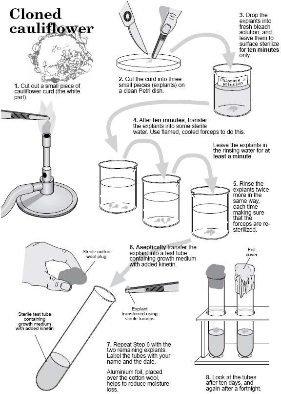
81 EXPERIMENT 3 74
82 Experiment 4: Callus Formation and Multiplication Background Callus is defined as an unorganized tissue mass growing on solid substrate. Callus forms naturally on plants in response to wounding, infestations, or at graft unions. Since extensive callus formation can be induced by elevated hormone levels, tissue culture media designed to produce callus contain pharmacological additions of cytokinins and auxins. Callus formation is central to many investigative and applied tissue culture procedures. Callus can be multiplied and later used to clone numerous whole plants. Additionally, various genetic engineering protocols employ callus initiation procedures after DNA has been inserted into cells; transgenic plants are then regenerated from transformed callus. In other protocols callus is generated for use in biotechnological procedures such as the formation of suspension cultures from which valuable plant products can be harvested. Callus Formation Explants from several parts of large intact plants can be used to form callus. The most successful explants are often young tissues of one or a few cell types. Pith cells of young stem are usually a good source of explant material. Initially, callus cells proliferate without differentiating, but eventually differentiation occurs within the tissue mass. Actively dividing cells are those uppermost and peripheral in the callus. The extent of overall differentiation usually depends on the hormone balance of the support medium and the physiological state of the tissue. EXPERIMENT 4 75
83 Callus Multiplication Actively growing callus can be initiated on culture media with an even physiological balance of cytokinin and auxin. (Callus Initiation Medium). After callus biomass increases two to four times (after 2 4 weeks of growth), callus can be divided and placed on fresh Callus Initiation Medium for callus multiplication. Multiplication procedures can be repeated several times (up to eight sequential transfers) before gross chromosome instability (or contamination) occurs. Differentiation and Plant Regeneration Multiplied callus can be stimulated to form shoots by increasing the cytokinin concentration and decreasing auxin content of culture media (Shoot Development Medium). Shoot masses can be cut apart and transferred to rooting medium. Once rooted, regenerated plants can be acclimatized to natural rather than "in vitro" growth conditions. Regenerated plants are especially valuable if the parent plant was itself unique or if the plants were genetically engineered. If, for example, multiplied callus was first used to form suspension cultures on which genetic engineering or cell selection was accomplished, resultant regenerated plants via tissue culture could possess special traits or capabilities. Materials and Methods Callus Formation 1. Obtain a 5-cm section of tobacco stem. 2. Cut off all leaves. 3. Immerse it in a beaker of 95% ethanol for 15 seconds. 4. In the laminar flow hood, expose the pith by cutting away epidermis, cortex, and vascular tissue with a sterile scalpel (Figure 4.1). 5. Slice the exposed length of pith into a sterile petri dish. EXPERIMENT 4 76
84 Cell and Tissue culture LAB MANUAL 6. Cover the dish to keep pith sterile. 7. Aseptically slice 5-mm cross-sections of pith. 8. Transfer one cross-section to each plate of Tobacco Callus Initiation Medium. 9. Cover the dishes, seal with parafilm, and place in an incubator at C. Callus Multiplication 1. Obtain a plate of tobacco callus. 2. Aseptically divide the callus into smaller pieces. 3. Transfer divided callus pieces to fresh Tobacco Callus Initiation Medium. Formulae for Tobaccoo Callus Initiationn Medium and Tobaccoo Shoot Development Medium Tobacco Callus Initiation Medium EXPERIMENT 4 77
85 Cell and Tissue culture LAB MANUAL Tobacco Shoot Development Medium Figure 4.1. By using pith explant and plating procedures, callus can be generated from pith cells placed on Tobacco Callus Initiation Medium. EXPERIMENT 4 78
86 Experiment 5: Studies on Carrot Callus Cultures Introduction: Objective: Apparatus Many crop plants offer no conventional method for vegetative propagation. One such species, the oil palm, is now being propagated on a commercial scale using a similar tissue culture technique to that employed in this investigation. Root explants are stimulated to produce undifferentiated callus by the provision of a suitable growth medium containing auxins. Eventually, embryoids spontaneously appear in the callus, and these are transferred to a medium which favors their proliferation. The shoots which develop are then transferred to a rooting medium and the whole plantlets, produced in this way, may then be hardened off and planted out. In this investigation, explants are obtained from the tap root of a carrot and stimulated to produce callus by the synthetic auxin 2,4-D. The callus cultures are maintained and subcultured on this medium and their properties are investigated. Embryoids that arise are observed microscopically or transferred to a hormone-free medium to encourage their further development. Each group will require: 1. Basic set of apparatus for aseptic handling: Sterile-handling cabinet, Bunsen burner, Beaker (400 ml) or stain jar + lid containing approx. 200 ml 70% ethanol solution (v/v). Beaker (1000 ml) or equivalent, ( waste EXPERIMENT 5 79
87 beaker), Sterile forceps, Sterile scalpel handle + blade, 70% ethanol solution (v/v), (200 ml approx.), Marker pen, Parafilm or insulating tape. 2. Additional set of apparatus: Tap root of carrot (Daucus carota), Scalpel (preferably sterilized), Sterile water (1 l approx.), preferably distilled Sterile petri dish (5), Sterile glass screw-top jar and lid, Sterile petri dish containing, growth medium (5), Bleach solution (250 ml approx.) + wetting agent. A) Initiation and Maintenance of Carrot Callus Cultures Notes on apparatus Procedure 1. The growth medium should be an agar medium containing mineral salts, sucrose, vitamins and 0.1 mg/ml, 2,4-D (2,4- dichlorophenoxyacetic acid). 2. The bleach solution + wetting agent can be either: a) 10% sodium chlorate(l) (hypochlorite) solution (v/v) (1.4% available chlorine) + 2 ml Teepol; or b) 20% solution (v/v) Domestos (1.4% available chlorine). You will be provided with a tap root of carrot (Daucus carota). If the tap root is undamaged and without internal air spaces, the interior of the root will be sterile. To eliminate tap roots with air spaces, only fresh, undamaged carrots which sink in water are used. Collect all your apparatus make up any solutions that you require and prepare your work-bench as suggested in the notes on aseptic handling. EXPERIMENT 5 80
88 1. Cut a section of tap root 3 6 cm long, discarding both ends of the root (Figure 5.1). Remove the epidermis and any blemishes with a scalpel, ensuring that you know which end was the root pole. Figure 5.1. Growth from explant after three weeks. The next part of the procedure is to sterilize the surface of the material by using a dilute solution of bleach. The solution must be handled with care and the following procedures must be carried out aseptically. 2. Place the tap-root section in a sterile, lidded jar and cover it with a chlorate(l) bleach solution (approx. 1.4% available chlorine) containing a wetting agent (detergent). Reseal the jar and shake for five seconds every five minutes for 20 minutes. 3. Pour off the bleach solution into the 1 l waste beaker. 4. The sterilized root is then washed four times by completely covering it with sterile water, resealing the jar, shaking for five seconds and pouring the water into the waste beaker. 5. Transfer the root to the base of a sterile petri dish, cut 1 cm from each end and discard this material. EXPERIMENT 5 81
89 6. Insert a sterile scalpel blade into the central core of xylem at the shoot (broader) end of the section; this will hold the material steady. 7. Cut 3 5 transverse sections, 1 3 mm thick, across the tap root and transfer each, shoot-pole uppermost, to a fresh sterile petri dish, which should be resealed after each operation. 8. Cut smaller sections, explants approximately 5 mm square, from each of these transverse sections by cutting across the cambium (Figure 5.2). The following method is recommended. i) Trim the cortex and some of the phloem from each transverse section (Figure 5.2b). ii) Cut off 5 mm-wide strips containing the cambium (Figure 5.2c). iii) Each strip can then be subdivided to produce 5 mm square explants, each containing parts of the phloem, xylem and cambium (Figure 5.2d). Figure 5.2. Production of tap root explants. EXPERIMENT 5 82

90 9. Transfer each explant, two per petri dish, root-pole downwards, to a complex sterile agar medium containing the hormone 2,4-D. 10. Seal each dish with Parafilm or insulating tape to reduce dehydration of the medium. Label each dish and incubate them upright, in the dark, at 25 C. 11. Examine your explants at weekly intervals. Record any changes that you observe. If your material is contaminated, you may transfer any undamaged explants to a fresh petri dish containing growth medium and incubate this as before (Steps 9 and 10). Maintaining growth of callus After 4 5 weeks, the callus produced will be well developed (Figure 5.3). To maintain its growth, it must be subcultured at regular 4-week intervals onto fresh medium. Unhealthy, dark necrotic tissue should be discarded. Figure 5.3. Well-developed carrot callus ready for subculture. EXPERIMENT 5 83
91 Apparatus Each group will require: 1. Basic set of apparatus for aseptic handling: 2. Additional set of apparatus: Sterile petri dish per callus to be subcultured, Petri dish (per callus) containing growth medium + 2,4-D Procedure Questions Prepare your work-bench as before. Carry out the following subculture procedure using aseptic handling techniques. 1. Transfer a well-developed callus to a sterile petri dish with sterile forceps. Reseal both dishes. 2. Barely raising the lid of the dish, use a sterile scalpel carefully to cut off healthy (yellow or cream-coloured) pieces of callus, approximately 5 mm cubes. 3. Transfer these callus cuttings to a petri dish containing freshgrowth medium, ensuring that the cuttings are widely spaced. 4. Seal the petri dish with tape or Parafilm and label it with the date and nature of the culture. Incubate the dish upright, in the dark, at 25 C. 1. Which tissues in the explant could have given rise to the callus produced? 2. Design and, if possible, carry out an experiment to eliminate one or more of these alternatives. EXPERIMENT 5 84
92 3. If the callus has not arisen from initially meristematic tissue, what does this suggest about the process of differentiation in plant tissues? B) Effect of Sucrose Concentration on Growth-Rate of Callus Cultures Apparatus Each group will require: 1. Basic set of apparatus for aseptic handling: 2. Additional set of apparatus: Procedure Carrot callus cultures, 3/4 days after subculture (3), *Balance, accurate to g *Sterile petri dish (3) Petri dish containing growth medium + 2,4-D + sucrose at a concentration in the range 0.75% 4% (w/v). *Required weekly 1. Transfer the petri dish with fresh medium to an incubator at 25 C. 2. Label clearly each of the empty petri dishes A, B, C, so that you can identify them. Weigh each of them accurately and record their masses in the form of a table. Carry out all of the following steps using aseptic handling techniques. Prepare your work-bench accordingly. When transferring callus, the lid of each dish should be barely lifted. 3. Using sterile forceps carefully transfer one callus culture to each of the sterile petri dishes. Try not to carry over any of the growth medium from the original culture. 4. Reweigh each of the dishes and their contents. Record the EXPERIMENT 5 85
93 new mass and determine the mass of each callus by difference. 5. Label the bottom of the petri dish containing growth medium, as shown in (Figure 5.4). 6. Using sterile forceps, transfer the callus from Dish A to the growth medium, placing it in position on the agar above the label A. Repeat this procedure with callus B and callus C. 7. Seal the dish with Parafilm or tape, and incubate it in the dark at 25 C. Figure 5.4. Label on base of petri dish containing growth medium. 8. Repeat Steps 1 7 at weekly intervals for six weeks, maintaining the code for each callus, A, B, C. 9. Express your results in graphical form and summarize the data in a simple table. EXPERIMENT 5 86
94 10. Collect summarized data from other members of your class. Figure 5.5. Biomass growth curve of a typical callus culture. Questions 1. Does the evidence suggest that the growth of frequently subcultured callus is arithmetical, logarithmic or of some other kind? (If necessary, plot a further graph of log10 biomass against time.). 2. (a). Determine the mean time taken for the callus mass to double for each sucrose concentration and plot a graph of mean doubling time against sucrose concentration. (b). Describe and explain the effects of varying sucrose concentration upon the growth of the callus. 3. (a). A callus culture that is maintained without transfer to fresh medium exhibits the growth curve EXPERIMENT 5 87
95 shown in Figure 5. Which phase of the growth curve most nearly resembles the pattern of growth that you observed? (b). Explain the patterns of growth observed in Phases B, C and D in the light of your experimental results. What other explanations are there for the shape of this curve? 4. Why was it suggested that the fresh growth medium should be incubated before the callus was transplanted? C) Embryogenesis in Carrot Callus Cultures Apparatus After eight weeks of growth on media containing 2,4-D, carrot callus cultures sometimes undergo developmental changes. 2,4-D stimulates disorganized growth but, under the low power of a microscope, organized structures may be observed at the edges of the callus. These putative embryoids may, if transplanted onto a hormone-free growth medium, give rise to shoots, roots and, ultimately, complete plants. Each group will require: 1. Basic set of apparatus for aseptic handling: 2. Additional set of apparatus: Sterile petri dish, Petri dish containing sterile, hormone-free growth medium (2), Microscope, slides and coverslips, Healthy carrot callus culture at least eight weeks old Glass rods. EXPERIMENT 5 88
96 Procedure The following procedure can be employed, as circumstances permit, at approximately weekly intervals after the eighth week of culture. Collect all your apparatus and prepare your work-bench as before. Carry out Steps 1 5 using aseptic handling techniques. 1. With sterile forceps, transfer a callus to a sterile petri dish. Reseal both dishes. 2. Barely raising the lid of the dish, use a sterile scalpel to cut off healthy (yellow or cream-coloured) pieces of callus, approximately 5 mm cubes, from the edge of the callus. 3. Transfer several cubes of callus to a petri dish containing hormone-free growth medium. Ensure that the cubes are widely spaced and pressed firmly onto the agar. Retain some cubes for Step Seal each dish with Parafilm or tape to reduce water loss. Label each dish with your name, date, details of the original callus and the medium s composition. 5. Incubate each dish upright, in the light, at 25 C. Examine your cultures at weekly intervals, recording any observations. Observation of embryoids 6. Remove and discard the dark, necrotic callus tissue. 7. Add 2 ml of water to the petri dish and, using a glass rod; gently break up as much as possible the remaining cubes of callus. 8. Transfer a drop of the resulting suspension to a clean microscope slide and cover with a coverslip. 9. (a). Examine the suspension, initially under low power. Typical embryoid structures are shown in Figure 6a 6d. (b) Draw any structures that you observe. EXPERIMENT 5 89
97 Figure 5.6a. Globular stage in embryoid development. Figure 5.6b. Heart-shaped stage. Figure 5.6c. Torpedo stage. Figure 5.6d. Late torpedo, early cotyledonary stage. Questions 1. Does your investigation support the statement that the nuclei of plant cells are totipotent? EXPERIMENT 5 90
98 D) Cytodifferentiation in Carrot Callus Cultures Apparatus Under the influence of the synthetic auxin 2,4-D, carrot explants grow to form a largely undifferentiated mass of cells, a callus. However, it is possible to observe differentiated xylem tracheary elements within the callus mass. Increased production of xylem (xylogenesis) may be stimulated in cultures from explants of entirely parenchymatous tissue by providing a particular balance of auxins and cytokinins in the medium. Each group will require: 1. Basic set of apparatus for aseptic handling: 2. Additional set of apparatus: Clean petri dish, Mounted needle (2), Forceps, Scalpel, Phloroglucinol + concentrated hydrochloric acid, Carrot callus (a subculture aged approx. three weeks), Microscope, slides and coverslips. Procedure 1. With forceps, remove the callus to a clean petri dish. 2. Cut off a small piece of healthy tissue (a 2 mm cube approx.) from the edge of the callus and transfer it to a clean microscope slide. 3. Add a small drop of water and macerate the tissue thoroughly with the mounted needles. 4. Add two drops of phloroglucinol and leave for 10 minutes, then drain off the liquid. EXPERIMENT 5 91
. Questions 1.")
99 5. Add one drop of concentrated hydrochloric acid. 6. Cover the material with a coverslip and observe, at first under the low power of a microscope. Lignin will be stained red/magenta. 7. Draw and label any lignified cells that you observe. Figure 5.7. Lignified structures from a carrot callus culture ( 250). Questions 1. When demonstrating cytodifferentiation in callus cultures, why is it important that the material used is a subculture rather than a primary callus grown from an explant? 2. Can you identify in your specimens any other differentiated cells? EXPERIMENT 5 92
100 E) Nuclear Division in Callus Cultures Apparatus Procedure Each group will require: Actively growing callus culture of carrot (Daucus carota), Scalpel, Boiling tube containing 5 ml 1M hydrochloric acid, Water bath at 60 C or beaker (400 ml) half full of water at 65 C, Petri dish, Forceps, Mounted needle (2), Microscope slides and coverslips, Aceto orcein stain (freshly, prepared and filtered), Bunsen burner, Microscope (low power objectives and magnification at least 250). 1. Place the boiling tube of acid into the water bath to warm up while you are carrying out Step Transfer the callus to a petri dish lid and, using a scalpel, cut portions of active material from the pale, creamy-white outer parts of the callus. 3. Using forceps, transfer the callus fragments to the boiling tube of acid, and shake gently. Return the tube to the water bath and allow the material to hydrolyse for 10 minutes. 4. After 10 minutes, carefully decant most of the acid and transfer the hydrolysed callus fragments to a petri dish base. 5. Use a scalpel to cut sections approximately 1 mm thick from fragments and place each section on a microscope slide. 6. Tease the fragment apart with the mounted needles. 7. Add two drops of aceto orcein stain and warm the slide gently until the stain steams, but does not boil. (Note: If the back of the slide is painful to the touch, then you are overheating it!) 8. Leave the material for 10 minutes to take up the stain. Carefully absorb excess stain with the edge of a piece of blotting paper. EXPERIMENT 5 93
101 Question 9. Add one drop of fresh stain, apply a coverslip, and press down hard through several thicknesses of blotting paper. Warm again gently. 10. Observe the material under low power and then at a higher magnification. Nuclear material should be densely stained and, in actively dividing cells, chromosomes should be visible as heavily-stained rod-like structures within the cells. 11. Draw any patterns that you observe and identify the phases of mitosis that they represent. 1. Of the cells with nuclei, what proportions are undergoing mitosis? EXPERIMENT 5 94
102 Experiment 6: Demonstration of "in vitro" Morphogenesis and Totipotency of Seedling Explants Introduction: This experiment gives a simple exercise demonstrating plant totipotency as well as the nutritional requirements of different plant organs employs shoot tip and root tip explants cut from aseptically germinated seedlings. Each type of explant (excised part of the intact organism) is transferred to three simple tissue culture media. Background Objective: During seed formation, the developing embryo and associated tissues tend to exclude pathogens and foreign materials that may be in the parent plant. Contents of the seed, then, are essentially aseptic and the resultant seedlings can be maintained in the aseptic condition if the outer surface of the seed (seed coat) is sterilized with sodium hypochlorite (or other surface sterilant) prior to germinating the seeds in a sterile petri dish. Students will sterilize seeds, maintain them in culture and develop plantlets to demonstrate plant totipotency as well as the nutritional requirements of different plant organs. Procedure 1. Collect all the apparatus that you will need and prepare any solutions that are required. EXPERIMENT 6 95
103 Procedure Methods: Week 1 The manipulations that are required for the germination of aseptic seedlings are outlined below and illustrated in Figure Figure 6.1. The manipulations required for the transfer of seedling explants to Mineral Salts (M) and Minimal Organic (O) growth media (Experiment 1, Methods: Week 2). EXPERIMENT 6 96
104 1. Outside the laminar flow hood: Place several (5 to 10) tomato or lettuce seeds in a small petri dish. Fill the petri dish with a 7% chlorox solution to which a drop of wetting agent has been added. The soapy chlorox solution is usually a good surface sterilant. Swirl the seeds intermittently during the 10- or 15-minute chlorox treatment. 2. Preparation for aseptic transfers: Begin by washing your hands and forearms with soap, followed by swabbing with 70% ethyl alcohol (EtOH). Sterilize the laminar flow hood by wiping the inside (top, sides, and bottom) with EtOH. Turn on the hood; minute operation of the hood before use insures aseptic conditions within the work area of the hood. Continue to swirl the seeds intermittently during the chlorox treatment. Prior to actual aseptic transfers inside the chamber, swab hands and forearms with EtOH again; also wipe the external surface of the petri dish before placing it inside the hood. The hood should contain the following: a large jar which can be used as a "sink," flasks of sterile water, forceps in a beaker of ethanol, sterile filter paper (5 7 cm diameter filter paper can be sterilized in glass petri dishes), and sterile petri dishes which can be used as the seed germination dishes. 3. Inside the laminar flow hood: Decant chlorox and replace with sterile H2O. Rinse this way twice. Each rinse should rest 10 minutes. Prepare the sterile germinating petri dish by retrieving a forceps from the 70% EtOH beaker. Using the sterile forceps remove three (3) rounds of sterile filter paper from a sterile container and place them in the germinating dish (sterile plastic petri dish). Finally, add 5 10 ml of sterile H2O to the seeds; decant seeds and water into the sterile germinating dish and incubate at 25 C until the next laboratory. (Both tomato and lightinsensitive lettuce seeds germinate in the light. Since shoots EXPERIMENT 6 97
105 become green but roots remain white under these conditions, seedling morphology is recognized more easily when lightgerminated.) Methods: Week 2 Examine the contents of the aseptic germinating dish without opening the lid. If there is no fungal or bacterial contamination around the seedlings, proceed; if contamination exists, request a dish of aseptic seedlings from the instructor. The manipulation required for the transfer of seedling explants to Mineral Salts (M) and Minimal Organic (O) growth media are outlined below and illustrated in Figure Swab chamber, hands, and upper/lower surfaces of petri dish with 70% ethanol. 2. Place germinating dish in transfer chamber. 3. Remove scalpel or scissors from the ETOH beaker already in the hood. Slip instrument between sheets of sterile toweling to remove ETOH (ethanol). 4. Lift one edge of lid and cut off no more than 10 mm of root tip. Excise two root tips. Lower lid. Place scalpel back into ETOH beaker. 5. Place tubes with sterile media into the transfer chamber. Use one tube of Minimal Organic Medium (O) and one of Mineral Salts Medium (M). Loosen these caps. 6. Remove forceps or inoculating loop from ETOH. Slip between sterile towelings to remove ETOH. 7. Remove excised root tip from germinating dish and transfer to the surface of the Minimal Organic Medium (O). Transfer second root tip to the surface of the Mineral Salts Medium (M). Caution: pick up root tip by the severed end; damage to the apical meristem disrupts mitosis! Measure or estimate length of root tips. Record. 8. Using aseptic technique as above, prepare and transfer one shoot tip into each type of media. Pick up the shoot tip by the severed end and insert it part way into the medium EXPERIMENT 6 98
106 with an overall vertical orientation of the cotyledons and shoot tip. Record size and shape of shoot tip. 9. Place the four tubes in a slant rack under lights. 10. Examine cultures each week. Record observations on the amount of growth and morphogenesis of both root and shoot cultures. Figure 6.2. The manipulations required for the germination of aseptic seedlings (Methods: Week 1). EXPERIMENT 6 99
107 Media Formulae Used in the Culture of Aseptic Seedling Explants (Root and Shoot Tips) Murashige Minimal Organic Medium with Agar and Sucrose [O] Mineral Medium Murashige and Skoog Salt Base (MS) with Agar [M] EXPERIMENT 6 100
108 Observations As cultures progress it should be possible to correlate size/shape changes with the nutrient content of the medium. A third medium, the B-deficient Medium, contains the same mineral constituents as does the Mineral Salts Medium (M) and the same amount of sucrose as the Minimal Organic Medium (O), but is devoid of B vitamins. Thus, this medium is referred to as B-deficient (B). Shoot tips and root tips have been transferred to this demonstration medium. Growth on this medium can be evaluated and compared with growth on student experimental media M and O. The Mineral Salts Medium (M) is the basal growth medium, supplying essential mineral nutrients for autotrophic plant growth. Predict the resultant growth in each circumstance, then monitor the growth and development of root and shoot explants in each medium (M, O, and B) and evaluate the following (record observations in Table 6.1): 1. Effect of B-vitamins on: a) shoot growth (increase in size) and morphology (change of shape), and b) root growth and morphology. 2. Effect of organic medium containing sucrose on: a) shoot growth and morphology, and b) root growth and morphology. Optional: Media M, O, and B are set up with root tip and shoot tip explants in darkness. This set of samples can be observed along with those in the light to evaluate the effect of light as well as media contents on the growth and development of plant organs. A row labeled "etiolation" would be added to the bottom of Table 6.1. EXPERIMENT 6 101
109 B-deficient Demonstration Medium [ B ] Experimental observations should include the following: 1. Parameters: temperature; light quality, duration, and intensity. 2. Drawings to scale. 3. Gross measurements (length, biomass accumulation, extent of morphogenesis, totipotency, primordia, number of branches) after 1 week and 2 weeks. 4. Net changes (Table 6.1). 5. Does the irregular orientation of the shoot explant change the growth pattern? How can these observations be explained? EXPERIMENT 6 102
110 Table 6.1. Use blank table to record results from Experiment 1. Shoot Root Media Mineral B-deficient Organic Mineral B-deficient Organic Cap color White Red Blue White Red Blue Change in length Change in Biomass Extent of morphogenesis Totipontecy Primordia Number of Branches Questions 1. Are all portions of the seedling totipotent? 2. Is autotrophic and heterotrophic growth of different plant organs apparent? 3. How could the experimental design be changed to more completely evaluate B-vitamin and extrasucrose effects? 4. If B vitamins seem important to root growth and development, how are vitamins probably supplied to plant roots in intact plants? EXPERIMENT 6 103
111 Experiment 7: Effects of Hormone Balance on Explant Growth and Morphogenesis Background Plant hormones, like animal hormones, are relatively small molecules that are effective at low tissue concentrations. The two types of plant hormones used in this experiment are cytokinins and auxins. Cytokinins are derived from adenine and produce two immediate effects on undifferentiated cells: the stimulation of DNA synthesis and increased cell division. Cytokinins also produce a delayed response in undifferentiated tissue which is the formation of shoot primordia. Both naturally occurring cytokinins, such as zeatin and synthetic analogs, such as kinetin, demonstrate cytokinin effects (Figure 7.1). Although low tissue concentrations of cytokinins (e.g., M zeatin) have noticeable effects, higher concentrations are found in actively dividing tissues such as those of plant embryos and developing fruits. Auxins are indole or indole-like compounds that stimulate cell expansion, particularly cell elongation. Auxins also promote adventitious root development. Indoleacetic acid (IAA), a naturally occurring auxin, and napthaleneacetic acid (NAA), a synthetic auxin, are depicted in Figure 7.1. Only small amounts of auxin ( M) are required to demonstrate an IAA response and even smaller amounts of synthetic auxin (e.g., NAA) are required for a tissue response. The likely reason for potency of synthetic auxins is their stability in plant tissue (i.e., the enzymes and processes EXPERIMENT 7 104
112 that degrade IAA do not "recognize" synthetic auxins). Synthetic auxins, then, are more effective hormones that also last for an extended length of time. Furthermore, light influences the physiological activity of IAA while synthetic auxins are not as light sensitive. Plant hormones do not function in isolation within the plant body, but, instead, function in relation to each other. Hormone balance is apparently more important than the absolute concentration of any one hormone. Both cell division and cell expansion occur in actively dividing tissue, therefore cytokinin and auxin balance plays a role in the overall growth of plant tissue. Since hormone balance is presumably important to the overall effect on growth and morphological changes, then the hormone differentials in each of the experimental media (A, B, and C) should produce somewhat different effects on the growth and development of excised explants. Figure 7.1. Structural formulae of plant growth and differentiation hormones used in the current Experiment. EXPERIMENT 7 105
113 Source of Aseptic Explant Material During seed development the embryos are formed with a placenta-like interface of intervening tissues between parental vascular supply and the embryo proper. This circumstance depresses passive migration of most foreign bodies and microorganisms into the developing embryo. If the embryo which often develops aseptically is released from the seed by aseptic germination procedures, then aseptic seedlings result. Any part of the aseptic seedling can be used as "in vitro" experimental material. In this experiment three explant types will be used: hypocotyl (undeveloped lower stem), epicotyl (shoot apex), and cotyledons. Media Formulae Media A, B, C, D, and E each contain the same complement of minerals, that is, salt base as in Medium D. The effect of minerals alone on explant growth and development constitute "basal growth rate" against which the effects of other media constituents can be measured. Medium D, then, serves as the base-line control for endogenous growth. Medium E, containing both essential minerals plus sucrose, constitutes the organic and inorganic control which can be used as the base-line indicator of explant growth when both minerals and sucrose are supplied. In addition to the substrate, sucrose, Medium E contains two organic growth factors, inositol and thiamine, which promote sugar metabolism and general anabolic growth processes. Medium E also contains additional phosphate thereby matching the phosphate concentrations of the experimental media (A, B, C). The experimental media contain similar inorganic and organic complements, but differ in hormone content. Since cytokinins are derived from adenine, adenine sulfate has been added to each of the experimental media (A, B, C). EXPERIMENT 7 106
114 In addition, either kinetin or 2iP ([2-isopentenyl]-adenine), both of which are synthetic cytokinins having immediate hormone activity, are supplemental cytokinins in media A, B, and C. Of the three experimental media, Medium A contains the highest amount of active cytokinin (30 mg/liter), while media B and C contain much lower amounts (2 mg/liter and 1 mg/liter, respectively). Conversely, Medium A contains only a small amount of auxin (0.3 mg IAA/liter), while Medium B contains a higher amount (2 mgiaa/liter). Medium C contains the lowest absolute concentration of auxin (0.1 mg NAA/liter), but this synthetic auxin is more efficient in promoting cell expansion and root formation than the naturally occurring auxin, IAA. Medium C, then, may actually represent the formula with the highest physiological auxin activity. Since cytokinin/auxin balance is reportedly important to the final overall effect on growth and development, the results for each experimental media may be expected to differ. The balance represented by Medium A is decidedly skewed towards a high cytokinin/low auxin ratio. Medium B represents a more even distribution of cytokinin and auxin, while Medium C may have an effectively higher auxin than cytokinin ratio because of the "in vivo" stability of NAA as well as its effectiveness as an auxin. EXPERIMENT 7 107
115 Cell and Tissue culture LAB MANUAL Media Formulae Used in the Culture of Explants "in vitro" Aseptic Seedling Murashige Shoot Multiplication Medium [A] (plus added sucrose) Murashige Shoot Multiplication Medium [B] (plus added sucrose) EXPERIMENT 7 108
116 Cell and Tissue culture LAB MANUAL Murashige Shoot Multiplication Medium [C] (plus added sucrose) Murashige Shoot Multiplication Medium [C] (plus added sucrose) Murashige and Skoog Salt Base (MS) Medium (Control, inorganic) [D] EXPERIMENT 7 109
117 Murashige Minimal Organic Medium with NaH 2 PO 4.H 2 O and Sucrose Medium [E] (Control, organic and inorganic) Methods: Week 1 (Aseptic Seed Germination) Materials: Procedure: Cucumber or tomato seeds or any other plant seeds, 95% ethanol, sterile jars, sterile water, 25% chlorox, freshly prepared, sterile forceps or spatula 1. Sterilize seeds (cucumber) for 1 minute with 95% EtOH. 2. Rinse in sterile water 3. Sterilize in 25% bleach for 5 minutes and rinse three times with sterile water. 4. Transfer 10 seeds with a sterile forceps or spatula to a nutrient agar plate. 5. Incubate for 1 week (20 23 C). Methods: Week 2 Seedling explants (approximately 1 cm in length) of aseptically germinated seeds can be cut from the seedlings as EXPERIMENT 7 110
118 shown in Figure 7.2. Aseptic techniques including the use of the laminar flow hood are necessary to evaluate growth experiments in which nutrient rich media are used. An explant (hypocotyl, epicotyl, or cotyledon) should be placed on each of the following experimental media: A) Murashige Shoot Multiplication, Medium [A], B) Murashige Shoot Multiplication, Medium [B], and C) Murashige Shoot Multiplication, Medium [C], which contain different concentrations of growth hormones. For comparison, one explant of each type should be placed on each control media: D) Murashige and Skoog Salt Base, Medium [D], and E) Murashige Minimal Organic Medium + Sucrose with NaH2PO4 H20, Medium [E]. Seal the petri dishes with parafilm to prevent desiccation and incubate at low light intensity until next week. Record incubation conditions. EXPERIMENT 7 111
119 Figure 7.2. The transfer of explants to differential growth media using aseptic techniques (Methods: Week 2). Questions 1. What effect does a hormone balance that is applied pharmacologically "in vitro" have on seedling explant growth and morphogenesis? 2. Which media formulations produce callus? To what extent? On which explants? Were these results predictable? 3. What media produce anomalous growth rather than callus? EXPERIMENT 7 112
120 4. How is this explained? Are adventitious roots formed on all explants in Medium [C]? 5. Which explants are readily totipotent on control media (D, E)? 6. Is a wound response associated with observed totipotency? 7. Does the hormone balance of experimental media (A, B, C) influence the extent of totipotency? 8. Are the organs that form during organogenesis complete and apparently functional? 9. Do microscopic observations of cell size, dimension, and orientation facilitate the interpretation of hormone effects? 10. Do these explants grown in Medium A (high cytokinin, low auxin) show rapid cell division without cell expansion? 11. Are shoot primordia forming on the explants in Medium A? 12. Are callus cells disorganized and oversized? Additional Studies 13. How does explant orientation in the medium influence results? 14. If callused explants are placed on Medium [A], will shoot primordia develop? 15. Is it possible to generate an entire plant from a callused explant? 16. Are serial transfers and the multiplication of callus possible? On which media? 17. Is it possible to clone whole plants from multiplied callus? Under what conditions? EXPERIMENT 7 113
121 Experiment 8: Control of organogenesis in cultures of Nicotiana tabacum Background In this experiment, you will investigate the effects of various combinations of growth substances on the nature and rate of organogenesis (organ, i.e. root and/or shoot, production) by Nicotiana explants. Such investigations are clearly a necessary step in the development of a reliable technique for the production of plantlets from the disorganized material used for genetic manipulation and for propagation. The possible role of plant hormones in development is also illustrated. Materials Apparatus tobacco buds sharp pointed forceps, surgical scissors 95% ethanol petri dishes of culture medium (1/2 strength MS, 2% sucrose 0.8% agar, glutamine [800 mg/liter], serine [100 mg/liter]) Each group will require: 1. Basic set of apparatus for aseptic handling: Sterile-handling cabinet, Bunsen burner, Beaker (400 ml) or stain jar + lid containing approx. 200 ml 70% ethanol solution (v/v). Beaker (1000 ml) or equivalent, ( waste beaker), Sterile forceps, Sterile scalpel handle + blade, 70% ethanol solution (v/v), (200 ml approx.), Marker pen, Parafilm or insulating tape. EXPERIMENT 8 114
122 2. Additional set of apparatus: Sterile water, 1 l approx., Sterile glass screw-top jar, Sterile petri dishes of agar medium containing mineral salts, vitamins, sucrose and hormones, Sodium chlorate(i) (hypochlorite) bleach solution (250 ml approx.) + wetting agent, Seedlings of Nicotiana tabacum. Notes on apparatus Procedure 1. The chlorate(l) bleach solution + wetting agent can be either: a) 2% sodium chlorate(l) solution (v/v) (approx. 0.25% available chlorine) + 2 ml Teepol; or b) 4% Domestos solution (v/v) (approx. 0.25% available chlorine). You will be given a jar containing one or more seedlings of Nicotiana tabacum, and a number of sterile petri dishes, each containing a complex agar medium. The media provided contain various concentrations and combinations of an auxin (either 1 Naphthyl-Acetic Acid [NAA] or Indol 3yl- Acetic Acid [IAA]) and a cytokinin (kinetin). Collect all your apparatus make up any solutions that you will require and prepare your work-bench as suggested in the notes on aseptic handling. The following procedure must be carried out using aseptic handling techniques. 1. Using sterile forceps and a scalpel, remove the apical (upper) 7 10 cm of the stem of the seedling with its attached leaves and transfer the material to a sterile petri dish lid. If EXPERIMENT 8 115
123 the seedling has been aseptically germinated and grown, surface sterilization (Steps 2, 3 and 4) may be omitted. 2. Transfer the plant material to a sterile, lidded glass jar and cover it with the bleach solution. Reseal the jar and shake it for five seconds. Shake for five seconds once every minute for exactly 10 minutes (or for the period suggested by your teacher). 3. After 10 minutes, pour off the bleach solution and wash the specimen four times by completely covering it with sterile water, resealing the jar, shaking for five seconds and discarding the water into the waste beaker. 4. Transfer the specimen to a sterile petri dish and remove and discard the basal (lowest) 1 cm of the specimen. 5. Cut the specimen into sections, keeping only the internodes (Figure 8.1) and transferring each to a separate sterile petri dish. Cut each internode into 1 2 cm lengths. Figure 8.1. Select explant materials (sections x) from a surfacesterile seedling EXPERIMENT 8 116
124 6. Hold each length with sterile forceps and split it lengthwise with a sterile scalpel (Figure 8.2) to expose the pith and other internal stem tissue. These are your explants. Figure 8.2. Cut 1 cm explants from the internode material. The explants need not be equal halves. 7. Transfer two explants, exposed pith downwards, to each of the different agar-based media. (Use a minimum of three dishes of each type). 8. Seal each dish with Parafilm or tape to limit dehydration of the medium. Label each clearly and incubate them upright, in the light, at 26 C. 9. (a). Examine your explants at weekly intervals and record any changes you observe. Uncontaminated, healthy explants from dishes showing contamination can be transferred to fresh growth medium and re-incubated (Steps 7 and 8). (b) Count the numbers of roots or shoots occurring on the explants. (Note: A shoot appearing within one week of the initial incubation should be ignored). (c) Tabulate your data and plot graphs of the number of roots and the number of shoots against time. Questions Determine the mean and standard deviation of the number of roots and of the number of shoots produced by a 1 cm length of explant on each of the different growth media after three weeks. EXPERIMENT 8 117
.")
125 3. 2. Which proportions of auxin and cytokinin stimulate the production of roots? Which stimulate shoot production? (a). Which type of phytohormone would you expect to predominate in commercial rooting hormone preparations sold for use with stem cuttings? 5. (b). What properties, other than the ability to stimulate residual meristems to produce roots, should be possessed by such a commercial preparation? Why should shoots developing within one week of incubation be ignored when collecting your data? Figure 8.3a. Organogenesis by Nicotiana stem explants (roots, root hairs clearly visible). EXPERIMENT 8 118

126 Figure 8.3b. Organogenesis by Nicotiana stem explants (roots, root hairs clearly visible). Extension Work: Production of Plantlets Apparatus After four or five weeks, shoots will be sufficiently developed to be transplanted to a hormone-free growth medium. Each group will require: 1. Basic set of apparatus for aseptic handling: 2. Additional set of apparatus: EXPERIMENT 8 119

127 Procedure Sterile petri dish, Sterile petri dish containing hormone-free growth medium (2 or more), Pots of potting compost, Plastic bags. 1. Aseptically transfer a well-developed, shoot-producing specimen to a petri dish. Reseal both dishes. 2. Carefully separate four or more larger shoots from each other but retain some of the mass of tissue at the basal end of the shoot. 3. Aseptically transfer two shoots to each of the dishes of hormone-free medium, pressing the basal ends gently onto the surface. 4. Seal each dish with Parafilm or tape and incubate in the light at 20 C 28 C. Examine weekly. 5. When roots have developed, transfer the plants to small pots containing damp sterile potting compost. Cover each pot with a plastic bag to maintain high humidity and incubate the plantlets in the light at 20 C 28 C for two or more weeks. Figure 8.4. Nicotiana tabacum plantlet produced in seven weeks by tissue culture. EXPERIMENT 8 120
128 Experiment 9: Control of organogenesis in cultures of petals of Saintpaulia ionatha (African violet) Background Apparatus The ability of Saintpaulia to regenerate from cuttings is renowned. This experiment investigates the effects of various combinations of the auxin NAA (1-naphthylacetic acid) and the cytokinins, kinetin and BAP (6-benzylaminopurine) upon organogenesis in cultures of African violet petals. Clearly, such investigations are a necessary part of the development of a reliable protocol for the propagation of plants by tissue culture. Saintpaulia ionatha is one of the many species of ornamental plant that are commercially propagated in this way. Each group will require: 1. Basic set of apparatus for aseptic handling: Sterile-handling cabinet, Bunsen burner, Beaker (400 ml) or stain jar + lid containing approx. 200 ml 70% ethanol solution (v/v). Beaker (1000 ml) or equivalent, ( waste beaker), Sterile forceps, Sterile scalpel handle + blade, 70% ethanol solution (v/v), (200 ml approx.), Marker pen, Parafilm or insulating tape. EXPERIMENT 9 121
129 2. Additional set of apparatus: Sterile water, 1 l approx., Sterile glass screw-top jar, Sterile petri dishes of agar medium containing mineral salts, vitamins, sucrose and hormones, Sodium chlorate(i) (hypochlorite) bleach solution (250 ml approx.) + wetting agent, Seedlings of Nicotiana tabacum. Students notes ), Sterile petri dish (2), Sterile screw-top jar (250 ml approx.), Sterile water (1000 ml), 5% Teepol (detergent) solution in water (v/v) (100 ml approx.), Sodium chlorate(i) (hypochlorite) bleach solution (200 ml approx.) + wetting agent, Petri dish containing growth medium (6 or more), Flowering African violet (Saintpaulia ionatha) or two mature flowers. Notes on apparatus Procedure 1. The chlorate(l) bleach solution + wetting agent can be either: a) 7% sodium chlorate(l) (1% available chlorine) + 1% Teepol in water (v/v); or b) 14% Domestos in water (v/v) (1% available chlorine). 2. The petri dishes should contain 20 ml of a sterile, agarbased growth medium, including sucrose, vitamins, mineral salts and mixtures in differing proportions of an auxin (NAA) and a cytokinin. Collect all the apparatus you need and make up any solutions. Prepare your work-bench as suggested in the notes on aseptic handling techniques. 1. Using forceps transfer two mature inflorescences from the Saintpaulia plant to a clean screw-top jar. Hold the inflorescences by their pedicels. EXPERIMENT 9 122
130 The following procedures should be carried out using aseptic handling techniques. 2. Cover the flowers with 100 ml detergent solution to dewax the material. Reseal the jar and, shaking frequently, leave it for one minute. 3. Pour off the liquid into the waste beaker, using the jar s lid to prevent loss of the flowers. You must now sterilize the surface of the petals. 4. Cover the flowers with 200 ml of the chlorate(l) solution and reseal the jar. (Caution: The chlorate(i) solution is a strong bleach, so it should be handled with care). 5. Shake the jar for five seconds every minute for exactly five minutes. 6. After exactly five minutes, pour off the liquid into the waste beaker as before and quickly rinse the material four times as follows. 7. Cover the flowers with approximately 200 ml sterile water, reseal the jar and shake for five seconds. Pour the water into the waste beaker. The material may be left in the final rinse water in the sealed jar until required. 8. Transfer each flower to a separate sterile petri dish and replace the lids. 9. Prepare explants by using a sterile scalpel to cut approximately 1 cm 1 cm squares of material from the petals of one of the flowers (Flower A). 10. Use sterile forceps to transfer two explants of petal material to a petri dish containing one of the growth media provided. EXPERIMENT 9 123

131 The explants should be widely spaced and pressed gently onto the agar. Repeat this procedure for each of the different media, so that the same flower acts as the source of explants used for every medium. 11. Label each dish with your name, the date, the flower code, and the growth medium. 12. Repeat Stages 9, 10 and 11 using the other flower (B) as a source of explants. 13. Seal each petri dish with Parafilm or insulating tape to reduce water loss. 14. Incubate the dishes upright, in the light, at 26 C. 15. Examine the material at half-weekly intervals, recording and sketching any changes that occur. Figure 9.1. Organogenesis by petal explants of Saintpaulia ionatha. Callus bearing root and shoot primordia. EXPERIMENT 9 124


132 Figure 9.2. Callus with more developed roots. Figure 9.3. Callus with leafy shoots. EXPERIMENT 9 125
FERN MULTIPLICATION KIT
 FERN MULTIPLICATION KIT Product No. F34 India Contact 1 2 3 6 I 7...7 Product Number Kit Components Product Description 2 1 EA Box 1 Instruction Manual 1 C913/C21-10ea Culture Containers 1 F91-1ea Forceps,
AFRICAN VIOLET MULTIPLICATION KIT
 AFRICAN VIOLET MULTIPLICATION KIT Product No. A137 India Contact Revised May 2013 Page 1 TABLES OF CONTENTS KIT COMPONENTS... 2 MATERIALS REQUIRED BUT NOT PROVIDED... 2 INTRODUCTION... 3 STERILIZATION
The preferred carbohydrate in plant cell culture media is sucrose. Glucose and fructose may be substituted in some cases, glucose being as
 Introduction One of the most important factors governing the growth and morphogenesis of plant tissues in culture is the composition of the culture medium. The basic nutrient requirements of cultured plant
HOSTA MULTIPLICATION KIT
 HOSTA MULTIPLICATION KIT Product No. H411 PhytoTechnology Laboratories Mailing Address: P.O. Box 12205, Shawnee Mission, KS 66282-2205 Phone: 1-888-749-8682 (1-913-341-5343 Outside the USA & Canada) Fax:
TISSUE CULTURE II. Organogenesis. PlSc 300 LAB Learn tissue culture techniques that promote organ formation.
 76 TISSUE CULTURE II Organogenesis PlSc 300 LAB 11 REFERENCE: Text: 663 666; 706 712; 717 718. OBJECTIVES: 1. Learn tissue culture techniques that promote organ formation. 2. Practice making leaf and shoot
TERRESTRIAL ORCHID SEED SOWING KIT
 TERRESTRIAL ORCHID SEED SOWING KIT Product No. O788 PhytoTechnology Laboratories Mailing Address: P.O. Box 12205, Shawnee Mission, KS 66282-2205 Phone: 1-888-749-8682 (1-913-341-5343 Outside the USA &
Sunlight. Chlorophyll
 Plant Nutrition & Soil Fertility Richard Smith Vegetable Crop and Weed Science Farm Advisor University of California Cooperative Extension Monterey, Santa Cruz and San Benito Counties Plant Nutrition 1.
Figure 6. The type of oil palm explants used in these experiments. A. Leaf explant, B. Zygotic embryos explant, and C. Female flower explant
 22 MATERIALS AND METHODS Location and Time This work was carried out in the Laboratory of Plant Biotechnology and Tissue Culture, Faculty of Agriculture, Bogor Agricultural University. It was started from
Arabidopsis Growing Protocol-A General Guide Xiyan Li *
 Arabidopsis Growing Protocol-A General Guide Xiyan Li * Department of Genetics, Stanford University, Stanford, USA *For correspondence: lixiyan@stanford.edu [Abstract] Arabidopsis as the model organism
DEPARTMENT OF BOTANY Guru Ghasidas Vishwavidyalaya, Bilaspur B. Sc. V Semester LBC 503 (Plant Tissue Culture)
") DEPARTMENT OF BOTANY Guru Ghasidas Vishwavidyalaya, Bilaspur B. Sc. V Semester LBC 503 (Plant Tissue Culture) Section A Multiple choice questions / Define the following terms 1 10 = 10 1. (d) Agrobacterium
DEPARTMENT OF BOTANY Guru Ghasidas Vishwavidyalaya, Bilaspur B. Sc. V Semester LBC 503 (Plant Tissue Culture) Section A Multiple choice questions / Define the following terms 1 10 = 10 1. (d) Agrobacterium
Transformation Protocol for Tobacco Abbreviated
 Transformation Protocol for Tobacco Abbreviated Streak out bacteria onto YM 2-3 days 28 C Co cultivation on RMOP 3-11 days in dark Selection on RMOP-TCH Subculture every 2 weeks Plantlets onto MST-TCH
Title: Development of Micropropagation and Acclimation Protocols for the Commercialization of a New Bonsai Ornamaental Tree for the California Market.
 Title: Development of Micropropagation and Acclimation Protocols for the Commercialization of a New Bonsai Ornamaental Tree for the California Market. Authors: Dan E. Parfitt 1, Helen M. Chan 2, and Ali
BIOLOGY CLASS IX ARTIFICIAL VEGETATIVE PROPAGATION
 BIOLOGY CLASS IX ARTIFICIAL VEGETATIVE PROPAGATION MICROPROPAGATION This a technique of propagating plants by removing tissues from a parent plant and culturing them in a chemical medium. 1. A sterilized
Fertilizers. TheBasics. Whats in a Fertilizer? Why use Fertilizer? Nitrogen (N) Nitrogen (N) Its on the Label! Other sources of Nitrogen
 Nitrogen (N) Its on the Label! Other sources of Nitrogen") Why use Fertilizer? Fertilizers TheBasics Expectations Want the Golf Course look. Helps in recovery from stress. Mowing, Heat, and Activities. New Hybrids of grass seed perform better than the older varieties,
Why use Fertilizer? Fertilizers TheBasics Expectations Want the Golf Course look. Helps in recovery from stress. Mowing, Heat, and Activities. New Hybrids of grass seed perform better than the older varieties,
Anorganic Fertilizer. Lenny Sri Nopriani, SP.MP
 Anorganic Fertilizer Lenny Sri Nopriani, SP.MP Jargon Fertilizer Synthetic Fertilizers can be organic or inorganic Organic: from living material, contains C-C bonds; does not imply organic certification
Soils and Fertilizers. Leo Espinoza Soils Specialist
 Soils and Fertilizers Leo Espinoza Soils Specialist 2004 What is soil? A collection of organic and inorganic bodies on the earth s surface, modified by the surrounding environment or even made by humans,
Plant Tissue Culture. Dr. Alain Lemansour UAE University Date Palm Development Research Unit Dept.
 Plant Tissue Culture By Dr. Alain Lemansour UAE University Date Palm Development Research Unit Dept. What is it? Tissue culture is the term used for the process of growing cells artificially in the laboratory
Literature on micropropagation of awa (Piper
 Biotechnology Jan. 2003 BI-4 Micropropagation of Awa (Kava, Piper methysticum) J. Kunisaki, A. Araki, and Y. Sagawa Department of Tropical Plant and Soil Sciences Literature on micropropagation of awa
CHAPTER 2. IN VITRO REGENERATION OF Gerbera jamesonii Bolus Ex. Hook f. Previous research has proven that Gerbera jamesonii could successfully be
 CHAPTER 2 IN VITRO REGENERATION OF Gerbera jamesonii Bolus Ex. Hook f. 2.1 EXPERIMENTAL AIMS Previous research has proven that Gerbera jamesonii could successfully be propagated in vitro. Different types
2. What are the advantages and disadvantages of plant tissue culture?
 UNIT VI - PLANT TECHNOLOGIES Lesson 2: Plant Tissue Culture Competency/Objective: Explain the process of tissue culture. Study Questions References 1. What is plant tissue culture? 2. What are the advantages
Chapter 1 Introduction
 Chapter 1 Introduction Experimental systems based on plant cell and tissue culture are characterized by the use of isolated parts of, called ex, obtained from an intact plant body and kept on, or in a
Full Disclosure, I create and sell Sumo Cakes Bonsai Fertilizer Basics
 Full Disclosure, I create and sell Sumo Cakes www.sumocakes.com Bonsai Fertilizer Basics Where does one start when talking about fertilizers for bonsai? The one thing I do know is that there is no such
Crop Management Practices. By Simon Bedasie
 Crop Management Practices Fertigation By Simon Bedasie Observations of a Visiting Consultant Water and fertilizer management looked like bucket chemistry. Very basic techniques for water and fertilizer
Question Bank Vegetative Propagation
 Question Bank Vegetative Propagation 1. Define vegetative reproduction. Ans. Vegetative reproduction is the formation of a new individual from any vegetative part of the plant body. 2. Mention any two
Title: Enhancement of Bramble Production in the Southeastern U.S. Through Micropropagation, Virus Indexing, and Field Evaluation for Trueness to Type
 Title: Enhancement of Bramble Production in the Southeastern U.S. Through Micropropagation, Virus Indexing, and Field Evaluation for Trueness to Type Progress Report SRSFC Project #2006 01 Research Proposal
1. Potassium nitrate for efficient plant nutrition
 Potassium nitrate is a unique source of potassium by its nutritional value and its contribution to the health and yields of plants. Potassium nitrate features desirable chemical and physical properties,
GEOL 408/508 INTRODUCTORY SOILS
 GEOL 408/508 INTRODUCTORY SOILS Lecture = 3 hrs/week Lab = 3hrs/week Course = 4 credits Name/Define the Following 1. N 2 2. NH 3 3. NH + 4 4. NO - 2 5. NO - 3 6. protein 7. organics 8. organic matter 9.
Application of Plant Hormones
 Application of Plant Hormones Agriculturists all over the world have developed certain unusual methods by which they successfully cultivate the crop plants. It is only in recent year s plant physiologists
STOLLER ENTERPRISES, INC. World leader in crop nutrition Potato Production Challenge - Page 1 of 9
 Potato Production Challenge - Page 1 of 9 Potato Production Challenge 1. Seed Piece A high quality seed piece will have fewer stems two to three. The stem will have more vigor and less disease. The quality
Problem. Can paper mill sludge be used as a fertilizer for plants and does it change the ph of the
 Problem Can paper mill sludge be used as a fertilizer for plants and does it change the ph of the substrate it was used in? This problem was selected to learn if the chemical make-up of paper mill sludge
Soils of Palau. Diversity and Fertility. Palau Livestock Management Workshop March 23-25, Jonathan Deenik, PhD University of Hawaii
 Soils of Palau Diversity and Fertility Jonathan Deenik, PhD University of Hawaii Palau Livestock Management Workshop March 23-25, 2011 Outline Plant nutrition Soil fertility Soil composition Texture and
Unit 5: Soil - Stages of Soil formation
 Biology Form 3 Page 68 Ms. R. Buttigieg Unit 5: Soil - Stages of Soil formation Soil is the layer of material covering parts of the land, the home of many organisms and the area from which land plants
Monitoring Nutrition for Crops
 Monitoring Nutrition for Crops Electrical conductivity ph Alkalinity Cation exchange and Anion exchange capacities Krishna Nemali, Ph.D. Why should you monitor nutrients for plants? $$ Nutrient deficiencies
What s in Your Media? Analysis of media components for micronutrient content
 What s in Your Media? Analysis of media components for micronutrient content Internal Report for Young Plant Research Center Partners. Not for publication in part or full without permission of authors.
GREENHOUSE SCREENING PROTOCOL OF COMMON BEAN FOR NODULATION AND NITROGEN FIXATION
 GREENHOUSE SCREENING PROTOCOL OF COMMON BEAN FOR NODULATION AND NITROGEN FIXATION Materials needed: 2:1, v/v Perlite:vermiculite potting media Plastic containers Rhizobia inoculant, liquid or peat based
Title: Lecture 16 Soil Water and Nutrients Speaker: Teresa Koenig Created by: Teresa Koenig, Kim Kidwell. online.wsu.edu
 Title: Lecture 16 Soil Water and Nutrients Speaker: Teresa Koenig Created by: Teresa Koenig, Kim Kidwell online.wsu.edu Photos courtesy of Rich Koenig Photos courtesy of USDA NRCS Soil Water and Nutrients
Soil: We Can t Grow without it!
 Outline Soil: We Can t Grow without it! Clay Robinson PhD, CPSS, CCA Associate Professor of Soils Illinois State University Which nutrients do plants need? Fertilizers: Which and how much? Nutrients and
MICROPROPAGATION OF CHRYSANTHEMUM (CHRYSANTHEMUM MORIFOLIUM) USING SHOOT TIP AS EXPLANT
 USING SHOOT TIP AS EXPLANT") MICROPROPAGATION OF CHRYSANTHEMUM (CHRYSANTHEMUM MORIFOLIUM) USING SHOOT TIP AS EXPLANT R. Nalini Department of Biotechnology, Adhiparasakthi Agricultural College (Affiliated to TNAU) G. B. Nagar, Kalavai,
MICROPROPAGATION OF CHRYSANTHEMUM (CHRYSANTHEMUM MORIFOLIUM) USING SHOOT TIP AS EXPLANT R. Nalini Department of Biotechnology, Adhiparasakthi Agricultural College (Affiliated to TNAU) G. B. Nagar, Kalavai,
ACHIEVEMENT LEVEL DESCRIPTORS
 ACHIEVEMENT LEVEL DESCRIPTORS FOR THE PLANT SYSTEMS ASSESSMENT Please note: Students performing at the Meets Expectations level also meet all standards at the Approaches Expectations level, and students
Understanding the Balance
 Understanding the Balance Soil is the foundation that builds strong crops, plants, roots and turf. Micro Biology in the soil is an absolute must in order to grow anything properly.. The Soil must contain
In vitro regeneration capacity of the ornamental varieties related to the cultural media
 In vitro regeneration capacity of the ornamental varieties related to the cultural media Elena Alina Rovină 1 *, Mirela Călinescu 1, Catiţa Plopa, 1 Valentina Isac 1 1 Research Institute for Fruit Growing
Name: B3 PLANT DISEASE. Class: Question practice. Date: 41 minutes. Time: 41 marks. Marks: BIOLOGY ONLY. Comments: Page 1 of 18
 B3 PLANT DISEASE Question practice Name: Class: Date: Time: 4 minutes Marks: 4 marks Comments: BIOLOGY ONLY Page of 8 To be healthy, plants need the right amount of mineral ions from the soil. The diagram
TISSUE CULTURE AND EX-VITRO ACCLIMATION OF RHODODENDRON sp.
 Buletin USAMV-CN, 64/2007 (-) ISSN 1454-232 TISSUE CULTURE AND EX-VITRO ACCLIMATION OF RHODODENDRON sp. Clapa Doina, Al. Fira Fruit Research Station Cluj, 5 Horticultorilor Str. Horticultorilor nr.5, 400457
Understanding Soil Microbiology and Biochemistry
 Understanding Soil Microbiology and Biochemistry Understanding the microbiology and biochemistry of the soil is an important part of understanding how plants grow, but what are soil microbiology and soil
Rooting Hormones; Procedures for collecting, preparing and storing cuttings.
 Unit 2. Rooting Hormones; Procedures for collecting, preparing and storing cuttings. During this laboratory period, you will be introduced to the various rooting hormones available to propagate plants
High Carbon Wood Fly Ash as a Biochar Soil Amendment
 High Carbon Wood Fly Ash as a Biochar Soil Amendment Preliminary Assessment Completed by Northern Tilth June 2010 Executive Summary Resource Management, Inc. (RMI) recycles wood ash from biomass plants
STUDIES ON THE EXPLORATION OF BIOFERTILIZERS IN TEA
 STUDIES ON THE EXPLORATION OF BIOFERTILIZERS IN TEA Synopsis of the thesis to be presented in candidature for the award of the Ph.D. degree of the Calicut University By N. Tensingh Baliah Under the Guidance
Fertilizers and nutrient management for hops. Diane Brown, Michigan State University Extension
 Fertilizers and nutrient management for hops Diane Brown, Michigan State University Extension Pre plant nutrient management for hops Soil test! Correct major issues before planting ph 6.2 to 6.5 Lime season
Soluble Fertilisers 30.0% 29.0% - 1.0%
 Sangral Soluble Sinclair Sangral fertilisers are readily soluble in water to provide instantly available nutrients for root and foliar uptake. A range of products is available to provide the optimum feed
Effect of Soil Amendment with Dry and Wet Distillers Grains on Growth of Canola and Soil Properties
 Effect of Soil Amendment with Dry and Wet Distillers Grains on Growth of Canola and Soil Properties Peiyuan Qian, Jeff Schoenau, Tom King and Cory Fatteicher Department of Soil Science, University of Saskatchewan,
Plantlet Regeneration via Somatic Embryogenesis in Four Species of Crocus
 Plantlet Regeneration via Somatic Embryogenesis in Four Species of Crocus Roya Karamian Department of Biology, Faculty of Science Bu-Ali Sina University Hamadan Iran Keywords: meristems, micropropagation,
International Journal of Pharma and Bio Sciences DIRECT REGENERATION FROM EMBRYO CULTURES OF LYCOPERSICON ESCULENTUM MILL CV PUSA RUBY ABSTRACT
 Research Article Plant science International Journal of Pharma and Bio Sciences ISSN 0975-6299 DIRECT REGENERATION FROM EMBRYO CULTURES OF LYCOPERSICON ESCULENTUM MILL CV PUSA RUBY P. KARUNAKAR RAO Department
Class 3: Soil Sampling and Testing. Chris Thoreau
 Class 3: Soil Sampling and Testing Chris Thoreau What is Soil Testing? What are We Testing For? Soil Sampling Methodologies Soil Testing Interpreting Soil Test Results What is Soil Testing? Soil testing
LaMotte. Soil Test Kit Garden Guide.
 LaMotte Soil Test Kit Garden Guide www.lamotte.com Visual Symptoms of Element Deficiencies Mineral Needle Color Stem Appearance Roots Nitrogen deficiency Light or pale green color Slender, may be succulent
Summary and conclusion
 Summary and conclusion 6.1 Chemical sterilization of Explants Surface sterilization of explants collected from mature trees is necessary before initiation of any in vitro culture. In the present study
Know Your Soil Getting the Dirt on Your Dirt FWAA. Dr. Steve Petrie Director of Agronomic Services
 Know Your Soil Getting the Dirt on Your Dirt FWAA Dr. Steve Petrie Director of Agronomic Services Crop productivity Genetics Soil Temperature Environ ment Crop Yield Moisture Manage ment Take the time
Sandy, low CEC, irrigated soil Acidic ph High ph Cold soils Soil low in P content or available P
 Nutrient Nitrogen (N) Phosphorus (P) Potassium (K) Plant Response to Nutrient Influences color, shoot, shoot density, root, rhizome and stolon, high temperature stress, cold tolerance, drought resistance,
Plant Breeding and Propagation
 Plant Breeding and Propagation Outline Crop Plant Evolution Plant Breeding Sexually Compatible Germplasm Sexually Incompatible Germplasm - Bacterial Gene Cloning Transgenic Plants Seed Propagation Asexual
Soil. Acidic soils... 1/19/2014
 Soil Chapter 8 Acidic Soils & Salt Affected Soils Pages 229 262 About 2.5 billion acres of mostly cultivated lands are affected by soil salinity Treating ph problems is less difficult than managing and
Plant Regeneration via Organogenesis and Somatic Embryogenesis
 18 Plant Biotechnology: Practical Manual 2 Plant Regeneration via Organogenesis and Somatic Embryogenesis 2.1 BACKGROUND AND BASICS One can achieve plant regeneration in a test tube by using suitable culture
Soils and plant nutrients
 Soils and plant nutrients Soils and soil texture Soil texture triangle Plant nutrients Major nutrients Secondary nutrients Minor nutrients Exchange Capacity (CEC) Soil ph Soils Consists of a series of
Plant Nutrition & Fertilizers WMAMG 1/31/17
 Plant Nutrition & Fertilizers WMAMG 1/31/17 By Dawn Pettinelli UConn Soil Nutrient Analysis Lab dawn.pettinelli@uconn.edu SOIL-PLANT SYSTEM From: Extension.org Relationships between plants & soils evolved
Introduction. Plant growth regulators are the critical media components in determining the developmental pathway of the plant cells.
 Introduction Callus is defined as an unorganized tissue mass growing on solid substrate. Callus forms naturally on plants in response to wounding, infestations, or at graft unions (Bottino, 1981). Callus
Soils. Nutrients needed by plants 10/21/2013. Consists of a series of layers called. Soils consists of: Topsoil (A horizon) upper most layer
 upper most layer") Soils Consists of a series of layers called Topsoil (A horizon) upper most layer Soils consists of: Mineral particles determine soil texture Organic matter Air and Soil organisms Nutrients needed by plants
Soils Consists of a series of layers called Topsoil (A horizon) upper most layer Soils consists of: Mineral particles determine soil texture Organic matter Air and Soil organisms Nutrients needed by plants
MATERIALS AND METHODS
 Materials & Methods MATERIALS AND METHODS Plant Material In the preliminary experiments, 30 varieties of grain Sorghum viz., IS 15, IS 28, IS 720, IS 745, IS 8887, IS 3040, IS 3949, IS 81, IS 4100, IS
Reap The Business of Success
 Reap The Business of Success INTRODUCIN G 4 NEW ADDITIONS IN VESTIGE AGRI AGRI BIOFUNGICIDE AGRI BIOFUNGICIDE Agri-Biofungicide is unique formulation of systemic and contact organic fungicide It multiplies
ACTIVITY 2 How Does Your Garden Grow?
 ACTIVITY 2 How Does Your Garden Grow? Study Question: Activity: Curriculum Fit: What is the optimum level of plant nutrient application? In this lesson students will conduct an experiment that will examine
How your rose bush makes food
 How your rose bush makes food Your rose bush takes in carbon dioxide from the air, water and nutrients from the ground and makes sugars and starches (the source of energy to do that is sunlight and the
Agrobacterium-mediated Rice Transformation from Mature Seeds
 Agrobacterium-mediated Rice Transformation from Mature Seeds Kimberly Nelson-Vasilchik, Joel Hague, and Albert Kausch The Plant Biotechnology Laboratory University of Rhode Island 530 Liberty Lane West
Micropropagation of Sterile and Non-Flowering Nicotiana Lines
 2008 The Japan Mendel Society Cytologia 73(1): 9 13, 2008 Micropropagation of Sterile and Non-Flowering Nicotiana Lines Sarala K.*, Rao R. V. S., Murthy T. G. K. and Satyavani J. V. R. Central Tobacco
THE INVESTIGATION BEGINS!
 THE INVESTIGATION BEGINS! Each team will create 4 water and soil mixtures. The water in the mixtures will dissolve the chemicals responsible for the ph of the soil. A farmer needs good land to successfully
Original Papers Plant Tissue Culture Letters, 11(3), (1994) INouE**, Koji NoMuRA***, Seiko TAKAHASHi**, OsHIMA* and Kiyoshi MASUDA**
, (1994) INouE**, Koji NoMuRA***, Seiko TAKAHASHi**, OsHIMA* and Kiyoshi MASUDA**") Original Papers Plant Tissue Culture Letters, 11(3), 184-190 (1994) An Efficient Procedure for Plant Regeneration from Long-term-cultured Callus Tissue of Hybrid Poplar (Populus sieboldii Miq. X Populus
Original Papers Plant Tissue Culture Letters, 11(3), 184-190 (1994) An Efficient Procedure for Plant Regeneration from Long-term-cultured Callus Tissue of Hybrid Poplar (Populus sieboldii Miq. X Populus
Peters. Peters Professional and Peters Excel provide a wide selection of the highest quality water-soluble fertilisers available
 Professional and Excel provide a wide selection of the highest quality water-soluble fertilisers available Why choose Peters? 1. Peters takes into account water quality 2. Peters leads the way technically
Basic Botany Master Gardener and Horticulture Training. Mark Heitstuman. WSU Asotin and Garfield County Director January 9, 2018
 Basic Botany 2018 Master Gardener and Horticulture Training Mark Heitstuman WSU Asotin and Garfield County Director January 9, 2018 Topics we ll discuss in Chapter 1- Basic Botany Plant life cycles Internal
VETIVER PROPAGATION. Nurseries and Large Scale Propagation. Dr Paul Truong Veticon Consulting Brisbane, Australia
 VETIVER PROPAGATION Nurseries and Large Scale Propagation Dr Paul Truong Veticon Consulting Brisbane, Australia 1. INTRODUCTION The Vetiver Network promotes the use of sterile vetiver cultivar to avoid
application in propagating Butterfly attracting Plants
 Plant Tissue Culture and its application in propagating Butterfly attracting Plants Kodiswaran Kandasamy Tissue Culture Unit Forest Biotechnology Division FRIM kodiswaran@frim.gov.my Cratoxylon formosum
Getting the Most out of Your Strawberry Soil Test Report. General Information
 gardening.cornell.edu Getting the Most out of Your Strawberry Soil Test Report 1. Be certain your soil ph is in the optimal range. 2. Add a nitrogen (N) source at the recommended rate and time. 3. Add
Nutrient Management And Nutrient Cycling Raymond C. Ward, President Ward Laboratories, Inc Kearney, NE
 Nutrient Management And Nutrient Cycling Raymond C. Ward, President Ward Laboratories, Inc Kearney, NE www.rayward@wardlab.com Take Good Soil Samples to Evaluate Soil Fertility Status Take soils samples
PlSc 300 Plant Propagation. Final Exam (150 points) Spring 2010
 Spring 2010") 1 PlSc 300 Plant Propagation Final Exam (150 points) Spring 2010 Directions: You have two hours to complete this exam. Try to answer the following questions to the best of your ability. If you have any
1 PlSc 300 Plant Propagation Final Exam (150 points) Spring 2010 Directions: You have two hours to complete this exam. Try to answer the following questions to the best of your ability. If you have any
Turfgrass Fertility. Soil Test Reports. Why Soil Sample? Interpretation & Understanding
 Turfgrass Fertility Clint Waltz, Ph.D. Associate Professor The University of Georgia Soil Test Reports Interpretation & Understanding Why Soil Sample? Information Chemical way of estimating the nutrients
Using Fertilizers: Feeding plants. Lydia Clayton UAF Cooperative Extension Service Kenai Peninsula District
 Using Fertilizers: Feeding plants Lydia Clayton UAF Cooperative Extension Service Kenai Peninsula District What to know before you grow? Proper fertilization is essential for: Disease control Weed control
SOIL SAMPLING AND ANALYSIS
 E AZ1412 ARIZONA COOPERATIVE TENSION College of Agriculture and Life Sciences College of Agriculture and Life Sciences 07/06 SOIL SAMPLING AND ANALYSIS This publication provides information on techniques
Soil and Plant Basics 2016 EKS Grazing School September 20, 2016
 Soil and Plant Basics 2016 EKS Grazing School September 20, 2016 David G. Hallauer Meadowlark Extension District Agent, Crops & Soils/Horticulture (785) 863 2212 What Plants Need to Grow Sunlight Air Water
REVOLUTIONARY SUGAR BASED PLANT FOOD SYSTEM GROWING WITH SUGARS HAS NEVER BEEN EASIER
 REVOLUTIONARY SUGAR BASED PLANT FOOD SYSTEM EASY AS 1-2-3 DRIVE OUT WASTE WITH SIMPLIFIED SUGAR BASED PLANT FOOD PROGRAM Seed/Clones Vegetative Flower Reload Soil BASED ON USING A LIGHT SOIL GROW MEDIUM
B. C. D. B. C. D. B. C. D. B. C. D.
 Student Name: Teacher: Date: Test: 9_12 Agriculture AP41 - Horticulture I Test 1 Description: Unit C-Plant Physiology District: Wake County Form: 501 1. Water loss from leaf surface through evaporation
Soil Test Report. HOME GARDEN VEGETABLE GARDEN Analysis Results
 Analysis VEGETABLE GARDEN Analysis Results Result Account Number: 54321 Page: 1 of 6 Soil Test Rating Very Low Low Medium High Very High Organic Matter, % 4.6 Phosphorus, ppm P (Bray-1 Equiv.) 12 Potassium,
Keywords: Agarwood, satalum, resin, micropropagation, tok
 Application of Tissue Culture Techniques in Woody Species Conservation, Improvement and Development in Vietnam: Agarwood (Aquilaria crassna Pierre ex LeComte) via tip Culture Tran Van Minh Institute of
ORGANOGENESIS IN CHRYSANTHEMUM MORIFOLIUM RAMAT (CULTIVAR ROMICA ) CALLUS CULTURES SMARANDA VÂNTU
 CALLUS CULTURES SMARANDA VÂNTU") Analele ştiinţifice ale Universităţii Al. I. Cuza Iaşi Tomul LII, s. II a. Biologie vegetală, 006 ORGANOGENESIS IN CHRYSANTHEMUM MORIFOLIUM RAMAT (CULTIVAR ROMICA ) CALLUS CULTURES SMARANDA VÂNTU Abstract:
Analele ştiinţifice ale Universităţii Al. I. Cuza Iaşi Tomul LII, s. II a. Biologie vegetală, 006 ORGANOGENESIS IN CHRYSANTHEMUM MORIFOLIUM RAMAT (CULTIVAR ROMICA ) CALLUS CULTURES SMARANDA VÂNTU Abstract:
* Unless otherwise stated all procedures should be carried out aseptically using sterile solutions and equipment.
 TRANSFORMATION OF ARABIDOPSIS THALIANA From Dirk Valvekens et al.(dirk Valvekens, Marc Van Montagu, & Mieke Van Lijsebettens 1988. Proc. Natl. Acad Sci. 85, 5536-5540.), as used by Caroline Dean et al
PRELIMINARY CONSERVATION EFFORT ON RHIZOPHORA ANNAMALAYANA KATHIR., THE ONLY ENDEMIC MANGROVE TO INDIA, THROUGH IN VITRO METHOD
 J. Plant Develop. 20(2013): 57 61 PRELIMINARY CONSERVATION EFFORT ON RHIZOPHORA ANNAMALAYANA KATHIR., THE ONLY ENDEMIC MANGROVE TO INDIA, THROUGH IN VITRO METHOD KATHIRESAN Kandasamy 1, RAVINDER Singh
Unit D: ph of Soil. Lesson : Identifying the ph Changes in Soil
 Unit D: ph of Soil Lesson : Identifying the ph Changes in Soil 1 KEY TERMS Soil ph Acid Lime Requirement 2 I. The ph scale measures how acidic or basic a substance is. A. It ranges from 0 to 14. 1. A ph
STUDIES ON DATE PALM IN TISSUE CULTURE
 STUDIES ON DATE PALM IN TISSUE CULTURE By GEHAN MOHAMED YOUSRY SALAMA B.Sc. Agric. Sci. (Horticulture), Fac. Agric., Ain Shams Univ., 1990 M.Sc. Agric. Sci. (Ornamental Horticulture), Fac. Agric., Cairo
Low-Cost Alternatives for Conventional Tissue Culture Media
 International Journal of Current Microbiology and Applied Sciences ISSN: 2319-7706 Volume 7 Number 04 (2018) Journal homepage: http://www.ijcmas.com Original Research Article https://doi.org/10.20546/ijcmas.2018.704.288
Chapter 21. Micropropagation of Cordyline terminalis. Tui Ray, Prasenjit Saha, and Satyesh C. Roy. Abstract. 1. Introduction
 Chapter 21 Micropropagation of Cordyline terminalis Tui Ray, Prasenjit Saha, and Satyesh C. Roy Abstract This protocol describes an ef fi cient and rapid method for large-scale multiplication of Cordyline
NUTRITION FOR GREENHOUSE TOMATOES
 dlinnesota Flower Growers Bulletin - July, 99 Volume, Number NUTRITION FOR GREENHOUSE TOMATOES Richard J. McAvoy Extension Specklist - Greenhouse Crops University of Connecticut Reprinted from the Connecticut
Micro propagation of sugarcane (Saccharum officinarum L.) through auxiliary buds
 through auxiliary buds") Micro propagation of sugarcane (Saccharum officinarum L.) through auxiliary buds P S \Varakagoda, S Subasinghe, D L C Kumari and T S Neththikumara Department of Crop Science, Faculty of Agriculture, University
Micro propagation of sugarcane (Saccharum officinarum L.) through auxiliary buds P S \Varakagoda, S Subasinghe, D L C Kumari and T S Neththikumara Department of Crop Science, Faculty of Agriculture, University
THE ROLE OF TISSUE CULTURE IN THE AVOCADO PLANT IMPROVEMENT SCHEME
 South African Avocado Growers Association Yearbook 1984. 7:25-26 THE ROLE OF TISSUE CULTURE IN THE AVOCADO PLANT IMPROVEMENT SCHEME Review DOROTHEA D NEL AND JM KOTZÉ DEPT OF MICROBIOLOGY AND PLANT PATHOLOGY,
IN VITRO REGENERATION OF APICAL SHOOT EXPLANTS FROM IN SITU GROWN UV-B STRESSED OCIMUM SANCTUM L
 IN VITRO REGENERATION OF APICAL SHOOT EXPLANTS FROM IN SITU GROWN UV-B STRESSED OCIMUM SANCTUM L *Rajendiran K., Kokilavani V. and Murugananthan P. Department of Botany, K.M. Centre for Post Graduate Studies,
Soil Ecology Project. Alex Pace, Meredith Callis, Cate Wilson, Sarah Welch
 Soil Ecology Project Alex Pace, Meredith Callis, Cate Wilson, Sarah Welch Background As a final project in ninth grade biology we were asked to do a soil ecology project. This project was to be created
AVOCADO CALLUS AND BUD CULTURE
 Proc. Fla. State Hort. Soc. 96:181-182. 1983. AVOCADO CALLUS AND BUD CULTURE M. J. Young University of Florida, IF AS, Fruit Crops Department, Gainesville, FL 32611 Additional index words, tissue culture,